. 2023 Apr 14;18(4):e0283001.
doi: 10.1371/journal.pone.0283001. eCollection 2023.

Analytical validation of a multi-cancer early detection test with cancer signal origin using a cell-free DNA-based targeted methylation assay

Affiliations

Analytical validation of a multi-cancer early detection test with cancer signal origin using a cell-free DNA-based targeted methylation assay

Gregory E Alexander et al. PLoS One. .

Abstract

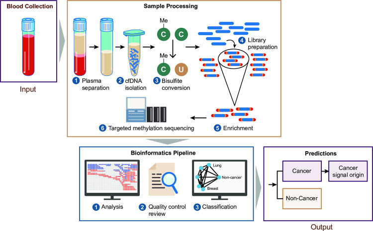
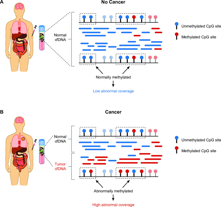
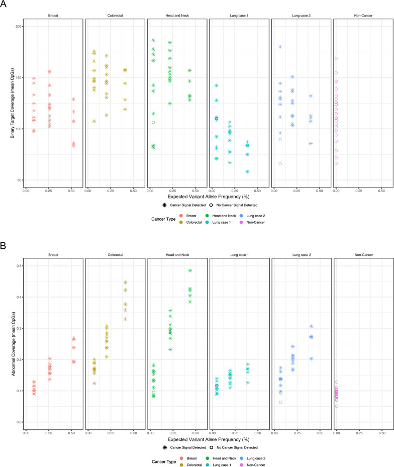
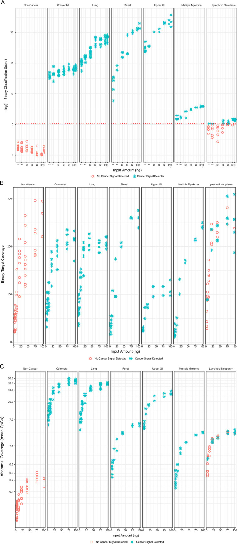
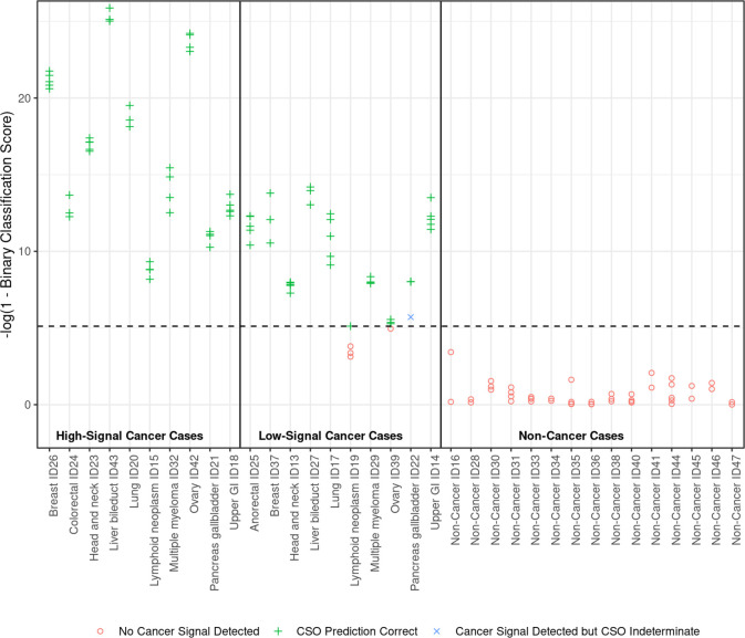
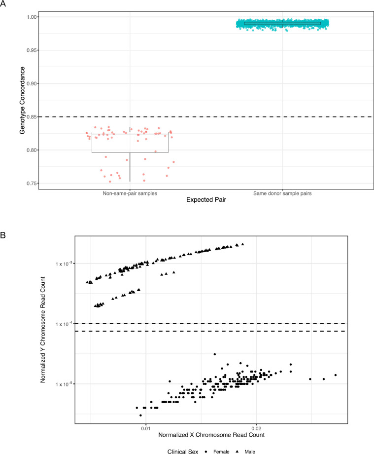
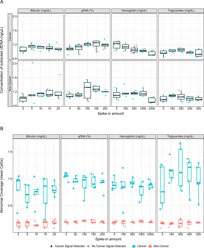
The analytical validation is reported for a targeted methylation-based cell-free DNA multi-cancer early detection test designed to detect cancer and predict the cancer signal origin (tissue of origin). A machine-learning classifier was used to analyze the methylation patterns of >105 genomic targets covering >1 million methylation sites. Analytical sensitivity (limit of detection [95% probability]) was characterized with respect to tumor content by expected variant allele frequency and was determined to be 0.07%-0.17% across five tumor cases and 0.51% for the lymphoid neoplasm case. Test specificity was 99.3% (95% confidence interval, 98.6-99.7%). In the reproducibility and repeatability study, results were consistent in 31/34 (91.2%) pairs with cancer and 17/17 (100%) pairs without cancer; between runs, results were concordant for 129/133 (97.0%) cancer and 37/37 (100%) non-cancer sample pairs. Across 3- to 100-ng input levels of cell-free DNA, cancer was detected in 157/182 (86.3%) cancer samples but not in any of the 62 non-cancer samples. In input titration tests, cancer signal origin was correctly predicted in all tumor samples detected as cancer. No cross-contamination events were observed. No potential interferent (hemoglobin, bilirubin, triglycerides, genomic DNA) affected performance. The results of this analytical validation study support continued clinical development of a targeted methylation cell-free DNA multi-cancer early detection test.


Conflict of interest statement

I have read the journal’s policy and the authors of this manuscript have the following competing interests: GA, WL, FEO, MR, PS, JRB, LE, GN, AM, PWM and NH are former employees of GRAIL, LLC and may have equity in the company. MHM is a consultant to Delfi Diagnostics and to Fellow Health and a former employee of GRAIL, LLC, with equity in all three companies. AMA is an advisor to Foresite Labs, San Francisco, California and Boston, Massachusetts, and a former employee of GRAIL, LLC, and holds equity in both companies. All other authors are employees of GRAIL, LLC, with equity in the company. GRAIL, LLC is a subsidiary of Illumina, Inc. currently held separate from Illumina Inc. under the terms of the Interim Measures Order of the European Commission dated 29 October 2021.

Figures

Fig 1
Fig 1. Overview of the multi-cancer early detection test workflow and computational pipeline.
Cell-free DNA (cfDNA) fragments are isolated from plasma and are treated with bisulfite to distinguish methylation patterns. Sequencing libraries are generated, are enriched for >100,000 genomic regions previously identified as having cancer- and/or tissue-specific methylation patterns, and then undergo targeted methylation sequencing. Following an initial analysis, if the data pass a quality-control review, a machine-learning classifier analyzes the targeted methylation sequencing data from cfDNA fragments to determine cancer status and, if cancer is detected, predict the cancer signal origin (16). C, cytosine; cfDNA, circulating cell-free DNA; Me, methyl group; U, uracil.
Fig 2
Fig 2. Abnormal coverage is a measure of abnormally methylated cell-free DNA (cfDNA) fragments analyzed by a targeted methylation-based multi-cancer early detection (MCED) test.
In a healthy individual (A), plasma contains normal cfDNA shed by normal cells. In an individual with cancer (B; lung tumor represented by orange circle), plasma contains a mixture of normal and tumor cfDNA. Cell-free DNA fragments contain CpG methylation sites that may be either unmethylated (blue lollipops) or methylated (red lollipops), which are reflected in cfDNA sequencing reads (blue or red segments). In tumor cfDNA, some methylation regions are abnormally methylated (dotted rectangles in B), unlike cfDNA from individuals without cancer (A). The representation (coverage) of these abnormally methylated regions is quantified by abnormal coverage, which is a measure of cancer detection in the MCED test. The coverage of other regions that are not affected by cancer (light blue and light red lollipops) is quantified by binary target coverage, which is a measure of baseline test performance. cfDNA, circulating cell-free DNA.
Fig 3
Fig 3. Binary target coverage is not correlated with variant allele frequency while abnormal coverage is correlated with variant allele frequency.
(A) Binary target coverage, which quantifies the coverage of methylation regions in cell-free DNA (cfDNA) fragments that are not affected by cancer, is a measure of the baseline performance of the multi-cancer early detection test. Binary target coverage is not affected by tumor content of admixtures (proportion of cfDNA fragments with variants identified in samples with matched tumor biopsy samples) in breast cancer (red), colorectal cancer (olive), head and neck cancer (green), and lung cancer (teal and blue) samples, similar to non-cancer samples (magenta). Samples with a cancer signal detected or not detected are indicated by asterisks or open circles, respectively. (B) Abnormal coverage is correlated with variant allele frequency (proportion of cfDNA fragments with variants identified in samples with matched tumor biopsy samples) in breast cancer (red), colorectal cancer (olive), head and neck cancer (green), and lung cancer (teal and blue) samples. Samples called as cancer or non-cancer are indicated by asterisks or open circles, respectively.
Fig 4
Fig 4
Correlations between cfDNA input amount, cancer type, and (A) binary classification score, (B) binary target coverage, and (C) abnormal coverage. Samples called as cancer or non-cancer are indicated by asterisks or open circles, respectively. (A) Binary classification score is positively correlated with the cell-free DNA input amount in the multi-cancer early detection test in colorectal, lung, renal, and upper gastrointestinal cancer and multiple myeloma. Scores for non-cancer samples are low and well below the detection cut-off (red dotted line). (B) Binary target coverage is positively correlated with the cell-free DNA input amount in the multi-cancer early detection test for both cancer (colorectal, lung, renal, and upper gastrointestinal [GI] cancer and multiple myeloma) and non-cancer samples. (C) Abnormal coverage is positively correlated (nonlinearly) with cfDNA input amount and generally lower in non-cancer samples than cancer samples. GI, gastrointestinal.
Fig 5
Fig 5. Binary classification scores of cancer and non-cancer samples were visually clustered in 3 groups.
The binary classification score of a sample is the percentile of its classifier-derived score among non-cancer samples in the training set. Binary classification scores were log transformed as shown on the y-axis to facilitate visualization. The horizontal dotted line indicates the threshold score used to call samples as cancer (indicated with a “+” if tissue of origin was predicted correctly or “×” if it was predicted as indeterminate) or non-cancer (red open circles). GI, gastrointestinal; ID, sample identifier.
Fig 6
Fig 6. Assessment of cross contamination in plasma samples with the cross-contamination module of the bioinformatics pipeline.
(A) Concordance of single-nucleotide polymorphism genotypes in same-donor sample pairs (teal circles) and non–same-pair samples (red circles). The horizontal dotted line indicates the threshold of 85% concordance used to call samples as not concordant (swapped) or concordant (not swapped). Boxes indicate 25th and 75th percentiles and the line inside corresponds to the median. Whiskers extend to minimum and maximum values, excluding outliers. Data points have been slightly offset horizontally (jittered) to better visualize points that may otherwise overlap. (B) Sex calls analysis. Male (triangles) and female (circles) samples were differentiated by the proportion of X and Y chromosome sequencing reads (x- and y-axes, respectively). The horizontal dotted lines indicate the thresholds used to call samples as male or female.
Fig 7
Fig 7
Effect of various concentrations of 4 potential interferents on (A) concentration of extracted cell-free DNA (cfDNA) and (B) abnormal coverage. Prior to cfDNA extraction, non-cancer plasma samples and cancer admixtures were spiked with bilirubin (0–20 mg/dL), high-molecular-weight genomic DNA (0–200% of total cfDNA extracted from unspiked samples), hemoglobin (0–2000 mg/dL), and triglycerides (0–500 mg/dL). Cancer admixtures were generated by adding abnormally methylated DNA from human HCT116 DKO cells to non-cancer plasma samples. Teal circles in Panel A represent samples. Boxes indicate 25th and 75th percentiles and the line inside corresponds to the median. Whiskers extend to minimum and maximum values, excluding outliers. gDNA, genomic DNA. Samples called as cancer or non-cancer are indicated by asterisks or open circles, respectively. Data points have been slightly offset horizontally (jittered) to better visualize points that may otherwise overlap.

Similar articles

Cited by

References

    1. Siu AL U. S. Preventive Services Task Force. Screening for breast cancer: U.S. Preventive Services Task Force recommendation statement. Ann Intern Med. 2016;164: 279–296. - PubMed
    1. U. S. Preventive Services Task Force, Curry SJ, Krist AH, Owens DK, Barry MJ, Caughey AB, et al. Screening for cervical cancer: US Preventive Services Task Force recommendation statement. JAMA. 2018;320: 674–686. doi: 10.1001/jama.2018.10897 - DOI - PubMed
    1. U. S. Preventive Services Task Force, Bibbins-Domingo K, Grossman DC, Curry SJ, Davidson KW, Epling JW Jr., et al. Screening for colorectal cancer: US Preventive Services Task Force recommendation statement. JAMA. 2016;315: 2564–2575. doi: 10.1001/jama.2016.5989 - DOI - PubMed
    1. Moyer VA U. S. Preventive Services Task Force. Screening for lung cancer: U.S. Preventive Services Task Force recommendation statement. Ann Intern Med. 2014;160: 330–338. - PubMed
    1. U. S. Preventive Services Task Force, Grossman DC, Curry SJ, Owens DK, Bibbins-Domingo K, Caughey AB, et al. Screening for prostate cancer: US Preventive Services Task Force recommendation statement. JAMA. 2018;319: 1901–1913. doi: 10.1001/jama.2018.3710 - DOI - PubMed

Publication types

MeSH terms