. 2023 Apr 14;9(15):eadf7586.
doi: 10.1126/sciadv.adf7586. Epub 2023 Apr 14.

Cryo-EM structure of the human Sirtuin 6-nucleosome complex

Affiliations

Cryo-EM structure of the human Sirtuin 6-nucleosome complex

Un Seng Chio et al. Sci Adv. .

Abstract

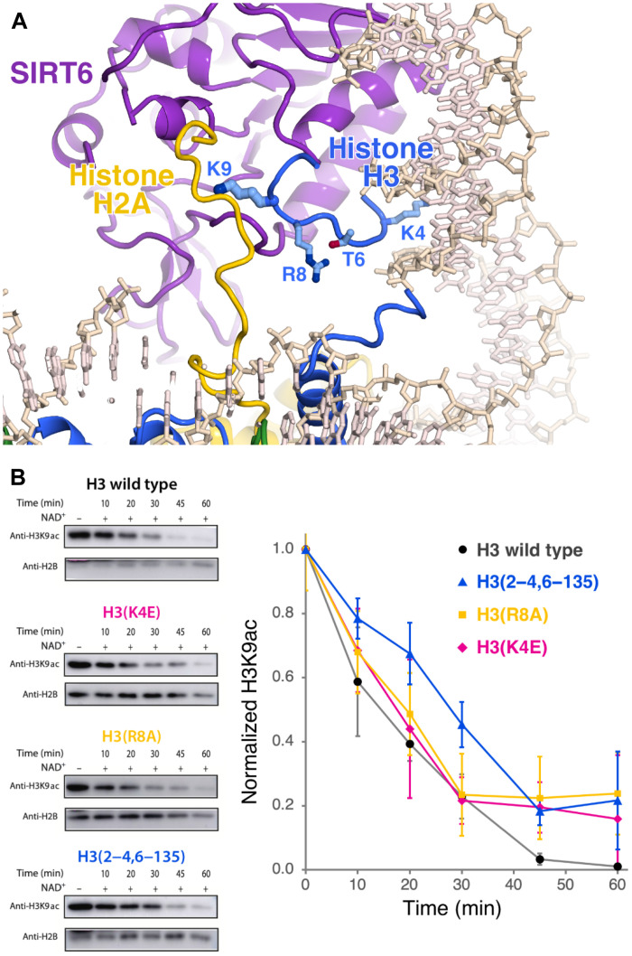
Sirtuin 6 (SIRT6) is a multifaceted protein deacetylase/deacylase and a major target for small-molecule modulators of longevity and cancer. In the context of chromatin, SIRT6 removes acetyl groups from histone H3 in nucleosomes, but the molecular basis for its nucleosomal substrate preference is unknown. Our cryo-electron microscopy structure of human SIRT6 in complex with the nucleosome shows that the catalytic domain of SIRT6 pries DNA from the nucleosomal entry-exit site and exposes the histone H3 N-terminal helix, while the SIRT6 zinc-binding domain binds to the histone acidic patch using an arginine anchor. In addition, SIRT6 forms an inhibitory interaction with the C-terminal tail of histone H2A. The structure provides insights into how SIRT6 can deacetylate both H3 K9 and H3 K56.


Figures

Fig. 1.
Fig. 1.. Overview of SIRT6 nucleosome structure.
(A) The 3.07-Å cryo-EM Coulomb potential density map of structure. (B) Cartoon representation of structure.
Fig. 2.
Fig. 2.. Interactions of SIRT6 zinc-binding domain with nucleosome acidic patch shown in cartoon representation.
Side chains of key residues are shown in stick representation.
Fig. 3.
Fig. 3.. Nucleosome binding of wild-type SIRT6 and SIRT6 containing point mutations in zinc-binding domain.
(A) Quantification of electrophoresis mobility shift nucleosome binding assay (EMSA) for wild-type (WT) and mutant SIRT6. (B) Results of time-resolved FRET nucleosome binding assay for wild type and mutant SIRT6. (C) Dissociation constants for wild-type and mutant SIRT6 determined by EMSA and time-resolved FRET. NA, not analyzed.
Fig. 4.
Fig. 4.. H3 K9Ac nucleosome deacetylation activity of wild-type SIRT6 and arginine anchor mutant SIRT6(R175A).
(A to C) Representative Western blots for the H3K9ac, H3K56ac, and H3K27ac deacetylation reactions for wild-type SIRT6 shown on top, and H3K9ac and H3K56ac deacetylation reactions for SIRT6(R175) shown at the bottom. (D to F) Corresponding plots for quantification of SIRT6-nucleosome deacetylation Western blots with SD error bars (n = 3).
Fig. 5.
Fig. 5.. Inhibitory interactions of histone H2A C-terminal tail near SIRT6 allosteric binding pocket.
(A) The histone H2A C-terminal binds to SIRT6 proximal to allosteric activators {MDL-801, modeled from two different structures [Protein Data Bank (PDB) 5Y2F and 6XV1]} and an allosteric inhibitor (catechin gallate, PDB 6QCJ). The modeled product analog, 2′-O-acyl-ADP-ribose, adopts a well-defined conformation in these three SIRT6-allosteric effector structures. The Cα positions of the H2A C-terminal residues 119 to 128 are shown as yellow spheres. Same color codes as for Fig. 2. (B) Deletion of the H2A C-terminal tail (residues 120 to 130) enhances SIRT6 nucleosomal H3K9ac deacetylase activity (normalized against histone H2B). Representative Western blot data (top) and plot for histone deacetylase (HDAC) assays with SD error bars shown (n = 3).
Fig. 6.
Fig. 6.. The SIRT6 catalytic domain interacts with nucleosomal DNA.
(A) Interactions of SIRT6 catalytic domain with nucleosomal DNA showing SIRT6 residues proximal to DNA. Same color code as for Fig. 2. (B) Quantification of electrophoresis mobility shift nucleosomal binding assay for wild-type (WT) and SIRT6 mutated in DNA binding residues.
Fig. 7.
Fig. 7.. SIRT6 nucleosomal substrate specificity.
(A) Cartoon and stick representation of SIRT6 binding of H3 tail around substrate residue K9. (B) Point mutations and deletions of H3 tail residues that interact with SIRT6 adversely affect SIRT6 nucleosomal histone H3K9Ac deacetylase activity. Representative Western blots are shown to the left and the plot for the HDAC assays is shown to the right with SD error bars (n = 3).
Fig. 8.
Fig. 8.. Cartoon and stick representation of SIRT6-nucleosome complex shows that histone H3 K56 is exposed but is at least 25 Å from H3 K9 and the SIRT6 catalytic site.
Same color code as for Fig. 2.

Update of

References

    1. R. H. Houtkooper, E. Pirinen, J. Auwerx, Sirtuins as regulators of metabolism and healthspan. Nat. Rev. Mol. Cell Biol. 13, 225–238 (2012). - PMC - PubMed
    1. Z. Mei, X. Zhang, J. Yi, J. Huang, J. He, Y. Tao, Sirtuins in metabolism, DNA repair and cancer. J. Exp. Clin. Cancer Res. 35, 182 (2016). - PMC - PubMed
    1. M. Kaeberlein, M. McVey, L. Guarente, The SIR2/3/4 complex and SIR2 alone promote longevity in Saccharomyces cerevisiae by two different mechanisms. Genes Dev. 13, 2570–2580 (1999). - PMC - PubMed
    1. R. Mostoslavsky, K. F. Chua, D. B. Lombard, W. W. Pang, M. R. Fischer, L. Gellon, P. Liu, G. Mostoslavsky, S. Franco, M. M. Murphy, K. D. Mills, P. Patel, J. T. Hsu, A. L. Hong, E. Ford, H. L. Cheng, C. Kennedy, N. Nunez, R. Bronson, D. Frendewey, W. Auerbach, D. Valenzuela, M. Karow, M. O. Hottiger, S. Hursting, J. C. Barrett, L. Guarente, R. Mulligan, B. Demple, G. D. Yancopoulos, F. W. Alt, Genomic instability and aging-like phenotype in the absence of mammalian SIRT6. Cell 124, 315–329 (2006). - PubMed
    1. Y. Kanfi, S. Naiman, G. Amir, V. Peshti, G. Zinman, L. Nahum, Z. Bar-Joseph, H. Y. Cohen, The sirtuin SIRT6 regulates lifespan in male mice. Nature 483, 218–221 (2012). - PubMed