Review
. 2023 Apr 13;186(8):1610-1626.
doi: 10.1016/j.cell.2023.03.010.

The role of extracellular vesicles in cancer

Affiliations
Review

The role of extracellular vesicles in cancer

Raghu Kalluri et al. Cell. .

Abstract

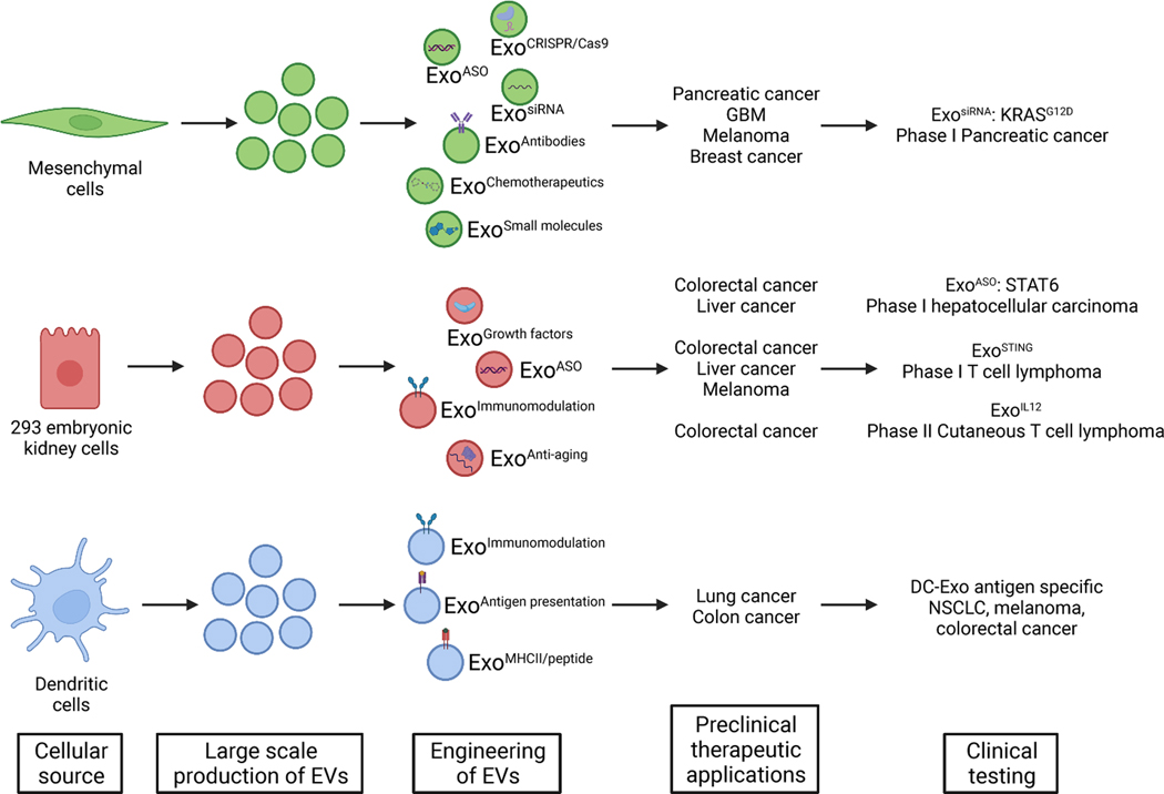
Intercellular communication is a key feature of cancer progression and metastasis. Extracellular vesicles (EVs) are generated by all cells, including cancer cells, and recent studies have identified EVs as key mediators of cell-cell communication via packaging and transfer of bioactive constituents to impact the biology and function of cancer cells and cells of the tumor microenvironment. Here, we review recent advances in understanding the functional contribution of EVs to cancer progression and metastasis, as cancer biomarkers, and the development of cancer therapeutics.


Conflict of interest statement

Declaration of interests MD Anderson Cancer Center and R.K. hold patents in the area of exosome biology and are licensed to Codiak Biosciences, Inc. MD Anderson Cancer Center and R.K. are stock equity holders in Codiak Biosciences, Inc. R.K. is a consultant and scientific adviser for Codiak Biosciences, Inc.

Figures

Figure 1.
Figure 1.. Extracellular vesicles include exosomes and ectosomes or microvesicles.
EVs present with a phospholipid bilayer membrane oriented similarly to that of the plasma membrane of the cell they are generated from. Exosomes are generated via the endosomal pathway, and result via the sequential invagination of the plasma membrane forming multivesicular bodies before they are released extracellularly. Ectosomes/microvesicles are generated via the outward budding of the plasma membrane. The mediators of different stages of multivesicular body formation, maturation, and release and RNA packaging into EVs are labeled. RBPs, RNA binding proteins. Created with BioRender.com.
Figure 2.
Figure 2.. Relative size and cargo of EVs.
(A) Average size of exosomes and microvesicles with respect to cellular components, including abundant proteins (albumin, hemoglobin, antibody), organelles (ribosomes, mitochondria), nucleotides and DNA, virus, as well as cellular byproducts: apoptotic bodies and platelets. (B) Composite cargo of exosomes, including surface receptors (protein, glycoprotein, glycans, ion channel receptors, G-protein coupled receptors, enzyme-linked receptors, integrins, etc.), transmembrane proteins (FasL, PD-L1, etc.), intracellular proteins, metabolites, lipids, and nucleic acids (RNA: mRNA, pre-/miRNA, piRNA, tRNA, snRNA, snoRNA, Y-RNA, circRNA; DNA: dsDNA, ssDNA, mtDNA, foreign DNA; cAMP). Created with BioRender.com.
Figure 3.
Figure 3.. Extracellular vesicles in metastatic disease.
Tumors release EVs, from both cancer cells and host cells of the tumor microenvironment (TME) into systemic circulation using both lymphatic and blood vessels. EVs interact with lymphoid organs including thymus and lymph nodes, with impact on T cell activation, DCs, and possibly aiding immune evasion. EVs also influence metastasis to lungs, liver, brain, and bone and possibly other non-metastatic sites by modifying vascular permeability and impacting immune cell recruitment, extracellular matrix (ECM) remodeling, and fibroblast activation. EVs exert their function by altering recipient cells via delivery of RNA, cytokines, chemokines, or growth factors, or surface protein signaling. Created with BioRender.com.
Figure 4.
Figure 4.. EVs as cancer biomarkers.
Cancer cells shed EVs with a characteristic cargo representing a range of cancer cell components, including nucleic acids, proteins, lipids, metabolites etc. EVs are found in all body fluids, including blood, urine, cerebrospinal fluids, saliva, sweat, tears, semen etc..) and may be enriched with various isolation protocols. EVs lend themselves to a multicomponent analysis reflecting a collection of cancer cells byproducts for biomarkers study, which likely offer a more comprehensive readout when compared to ctDNA analysis alone. Created with BioRender.com.
Figure 5.
Figure 5.. EVs as anti-cancer therapeutic agents.
Distinct cellular sources have been used to generate EVs in large scale for clinical trials. EVs engineering include the incorporation of a cargo (e.g. ASO, siRNA, chemotherapeutics etc), enriching for exosomes with unique surface protein presentation (e.g. antigen, immune modifying receptor). Preclinical studies in various tumor models and tumor types informed ongoing clinical trial design. EVs offer a novel therapeutic platform for cancer treatment, from personalized medicine to immunotherapy and targeted therapy with novel safety and efficacy profiles. Created with BioRender.com.

References

    1. Johnstone RM, Adam M, Hammond JR, Orr L, and Turbide C. (1987). Vesicle formation during reticulocyte maturation. Association of plasma membrane activities with released vesicles (exosomes). J Biol Chem 262, 9412–9420. - PubMed
    1. Pan BT, Teng K, Wu C, Adam M, and Johnstone RM (1985). Electron microscopic evidence for externalization of the transferrin receptor in vesicular form in sheep reticulocytes. J Cell Biol 101, 942–948. 10.1083/jcb.101.3.942. - DOI - PMC - PubMed
    1. Kugeratski FG, Hodge K, Lilla S, McAndrews KM, Zhou X, Hwang RF, Zanivan S, and Kalluri R. (2021). Quantitative proteomics identifies the core proteome of exosomes with syntenin-1 as the highest abundant protein and a putative universal biomarker. Nat Cell Biol 23, 631–641. 10.1038/s41556-021-00693-y. - DOI - PMC - PubMed
    1. Kowal J, Arras G, Colombo M, Jouve M, Morath JP, Primdal-Bengtson B, Dingli F, Loew D, Tkach M, and Thery C. (2016). Proteomic comparison defines novel markers to characterize heterogeneous populations of extracellular vesicle subtypes. Proc Natl Acad Sci U S A 113, E968–977. 10.1073/pnas.1521230113. - DOI - PMC - PubMed
    1. Mathieu M, Nevo N, Jouve M, Valenzuela JI, Maurin M, Verweij FJ, Palmulli R, Lankar D, Dingli F, Loew D, et al. (2021). Specificities of exosome versus small ectosome secretion revealed by live intracellular tracking of CD63 and CD9. Nat Commun 12, 4389. 10.1038/s41467-021-24384-2. - DOI - PMC - PubMed

Publication types

Substances