

Obliterated cavum septi pellucidi: Clinical significance and role of fetal magnetic resonance
- PMID: 37059118
- PMCID: PMC10201966
- DOI: 10.1111/aogs.14575
Obliterated cavum septi pellucidi: Clinical significance and role of fetal magnetic resonance
Abstract
Introduction: The objective of this study was to describe a cohort of fetuses with an ultrasound prenatal diagnosis of obliterated cavum septi pellucidi (oCSP) with the aim to explore the rate of associated malformations, the progression during pregnancy and the role of fetal magnetic resonance imaging (MRI).
Material and methods: This was a retrospective multicenter international study of fetuses diagnosed with oCSP in the second trimester with available fetal MRI and subsequent ultrasound and/or fetal MRI follow-up in the third trimester. Where available, postnatal data were collected to obtain information on neurodevelopment.
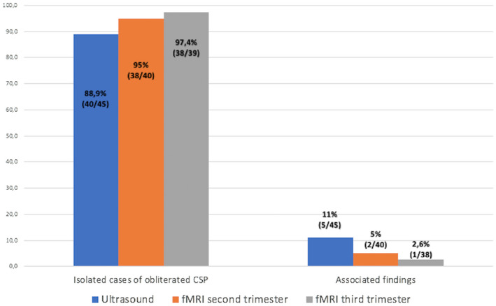
Results: We identified 45 fetuses with oCSP at 20.5 weeks (interquartile range 20.1-21.1). oCSP was apparently isolated at ultrasound in 89% (40/45) and fetal MRI found additional findings in 5% (2/40) of cases, including polymicrogyria and microencephaly. In the remaining 38 fetuses, fetal MRI found a variable amount of fluid in CSP in 74% (28/38) and no fluid in 26% (10/38). Ultrasound follow-up at or after 30 weeks confirmed the diagnosis of oCSP in 32% (12/38) while fluid was visible in 68% (26/38). At follow-up MRI, performed in eight pregnancies, there were periventricular cysts and delayed sulcation with persistent oCSP in one case. Among the remaining cases with normal follow-up ultrasound and fetal MRI findings, the postnatal outcome was normal in 89% of cases (33/37) and abnormal in 11% (4/37): two with isolated speech delay, and two with neurodevelopmental delay secondary to postnatal diagnosis of Noonan syndrome at 5 years in one case and microcephaly with delayed cortical maturation at 5 months in the other.
Conclusions: Apparently isolated oCSP at mid-pregnancy is a transient finding with the visualization of the fluid later in pregnancy in up to 70% of cases. At referral, associated defects can be found in around 11% of cases at ultrasound and 8% at fetal MRI indicating the need for a detailed evaluation by expert physicians when oCSP is suspected.
Keywords: cavum septi pellucidi; fetal brain; fetal magnetic resonance; neurosonography; postnatal neurodevelopmental outcome.
© 2023 The Authors. Acta Obstetricia et Gynecologica Scandinavica published by John Wiley & Sons Ltd on behalf of Nordic Federation of Societies of Obstetrics and Gynecology (NFOG).
Conflict of interest statement
None.
Figures


Similar articles
-
Obliterated cavum septi pellucidi: is it always a benign finding? A case report and narrative review of the literature.J Matern Fetal Neonatal Med. 2023 Dec;36(2):2232075. doi: 10.1080/14767058.2023.2232075. J Matern Fetal Neonatal Med. 2023. PMID: 37414745 Review.
-
Role of magnetic resonance imaging in fetuses with mild or moderate ventriculomegaly in the era of fetal neurosonography: systematic review and meta-analysis.Ultrasound Obstet Gynecol. 2019 Aug;54(2):164-171. doi: 10.1002/uog.20197. Epub 2019 Jul 11. Ultrasound Obstet Gynecol. 2019. PMID: 30549340
-
Long-term postnatal outcome of fetuses with prenatally suspected septo-optic dysplasia.Ultrasound Obstet Gynecol. 2020 Sep;56(3):371-377. doi: 10.1002/uog.22018. Ultrasound Obstet Gynecol. 2020. PMID: 32196785 Free PMC article.
-
What Are the Double Lines of the Fetal Cavum Septi Pellucidi on Ultrasound?J Ultrasound Med. 2022 Aug;41(8):1907-1914. doi: 10.1002/jum.15867. Epub 2021 Nov 9. J Ultrasound Med. 2022. PMID: 34751464
-
Spectrum of brain malformations in fetuses with mild tubulinopathy.Ultrasound Obstet Gynecol. 2023 Jun;61(6):740-748. doi: 10.1002/uog.26140. Ultrasound Obstet Gynecol. 2023. PMID: 36484554
References
-
- Malinger G, Paladini D, Haratz KK, Monteagudo A, Pilu GL, Timor‐Tritsch IE. ISUOG practice guidelines (updated): sonographic examination of the fetal central nervous system. Part 1: performance of screening examination and indications for targeted neurosonography. Ultrasound Obstet Gynecol. 2020;56:476‐484. - PubMed
-
- Society for Maternal‐Fetal Medicine (SMFM) , Ward A, Monteagudo A. Absent Cavum Septi Pellucidi. Am J Obstet Gynecol. 2020;223:23‐26. - PubMed
-
- Salomon LJ, Alfirevic Z, Berghella V, et al. ISUOG practice guidelines (updated): performance of the routine mid‐trimester fetal ultrasound scan. Ultrasound Obstet Gynecol. 2022;59:840‐856. - PubMed
-
- Jou HJ, Shyu MK, Wu SC, Chen SM, Su CH, Hsieh FJ. Ultrasound measurement of the fetal cavum septi pellucidi. Ultrasound Obstet Gynecol. 1998;12:419‐421. - PubMed
-
- Falco P, Gabrielli S, Visentin A, Perolo A, Pilu G, Bovicelli L. Transabdominal sonography of the cavum septum pellucidum in normal fetuses in the second and third trimesters of pregnancy. Ultrasound Obstet Gynecol. 2000;16:549‐553. - PubMed
Publication types
MeSH terms
LinkOut - more resources
Full Text Sources
