. 2023 Jun;22(6):100547.
doi: 10.1016/j.mcpro.2023.100547. Epub 2023 Apr 13.

Biomarkers Found in the Tumor Interstitial Fluid may Help Explain the Differential Behavior Among Keratinocyte Carcinomas

Affiliations

Biomarkers Found in the Tumor Interstitial Fluid may Help Explain the Differential Behavior Among Keratinocyte Carcinomas

Clara Matas-Nadal et al. Mol Cell Proteomics. 2023 Jun.

Abstract

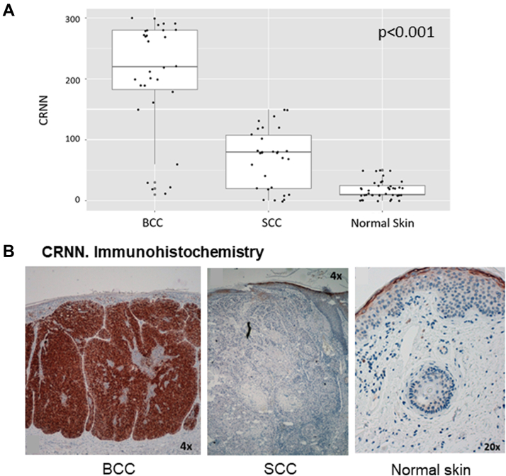
Basal cell carcinomas (BCCs) and cutaneous squamous cell carcinomas (SCCs) are the most frequent types of cancer, and both originate from the keratinocyte transformation, giving rise to the group of tumors called keratinocyte carcinomas (KCs). The invasive behavior is different in each group of KC and may be influenced by their tumor microenvironment. The principal aim of the study is to characterize the protein profile of the tumor interstitial fluid (TIF) of KC to evaluate changes in the microenvironment that could be associated with their different invasive and metastatic capabilities. We obtained TIF from 27 skin biopsies and conducted a label-free quantitative proteomic analysis comparing seven BCCs, 16 SCCs, and four normal skins. A total of 2945 proteins were identified, 511 of them quantified in more than half of the samples of each tumoral type. The proteomic analysis revealed differentially expressed TIF proteins that could explain the different metastatic behavior in both KCs. In detail, the SCC samples disclosed an enrichment of proteins related to cytoskeleton, such as Stratafin and Ladinin-1. Previous studies found their upregulation positively correlated with tumor progression. Furthermore, the TIF of SCC samples was enriched with the cytokines S100A8/S100A9. These cytokines influence the metastatic output in other tumors through the activation of NF-kB signaling. According to this, we observed a significant increase in nuclear NF-kB subunit p65 in SCCs but not in BCCs. In addition, the TIF of both tumors was enriched with proteins involved in the immune response, highlighting the relevance of this process in the composition of the tumor environment. Thus, the comparison of the TIF composition of both KCs provides the discovery of a new set of differential biomarkers. Among them, secreted cytokines such as S100A9 may help explain the higher aggressiveness of SCCs, while Cornulin is a specific biomarker for BCCs. Finally, the proteomic landscape of TIF provides key information on tumor growth and metastasis, which can contribute to the identification of clinically applicable biomarkers that may be used in the diagnosis of KC, as well as therapeutic targets.

Keywords: S100A9; basal cell carcinoma; keratinocyte carcinomas; proteomics; squamous cell carcinoma; tumor interstitial fluid.


Conflict of interest statement

Conflict of interest The authors declare no conflict of interest to disclosure.

Figures

None
Graphical abstract
Figure 1
Figure 1
Proteomic analysis of the tumor interstitial fluid from keratinocyte carcinomas.A, bar graphics representing the number of proteins recovered per sample. B, box diagram representing the proteins recovered per each type of sample. C, PCA graphics with the proteins quantified in at least 50% of the samples. D, heat map showing the protein profile of each sample. The expression levels of the proteins are stated by color changes. The highest expression is stated in red and the lowest is in green. PCA, principal component analysis.
Figure 2
Figure 2
Gene ontology cellular component of KC. Bar graphics showing the GO cellular component of SCC- (A) and BCC- (B) enriched proteins. BCC, basal cell carcinoma; GO, Gene Ontology; KC, keratinocyte carcinoma; SCC, squamous cell carcinoma.
Figure 3
Figure 3
The enrichment of the TIF samples from SCC, BCC, and NS was analyzed by immunoblot. Pictures of the gels stained with Coomassie Blue are shown as a loading control. BCC, basal cell carcinoma; SCC, squamous cell carcinoma; TIF, tumor interstitial fluid; NS, normal skin.
Figure 4
Figure 4
Cornulin expression in KCs and normal skin.A, box diagram showing CRNN expression in the neoplastic cells of BCC, SCC, and healthy skin according to the histoscore values [0–300]. Differences in the expression were statistically significant (p < 0.001) for the three types of samples. B, representative images of immunohistochemistry. CRNN expression in BCC, SCC, and normal skin. BCC, basal cell carcinoma; CNN, Cornulin; KC, keratinocyte carcinoma; SCC, squamous cell carcinoma.
Figure 5
Figure 5
Ladinin-1 expression in KCs and normal skin.A, box diagram showing LAD1 expression in the neoplastic cells of BCC, SCC, and healthy skin according to the histoscore values. Differences in the expression were statistically significant (p < 0.01) for the three types of samples. BD, representative images of immunohistochemistry. LAD1 expression in BCC (B), SCC (C), and normal skin (D). BCC, basal cell carcinoma; KC, keratinocyte carcinoma; SCC, squamous cell carcinoma.
Figure 6
Figure 6
S100A9 expression in KCs and normal skin.A, box diagram and IHC comparing S100A9 expression in the neoplastic cells of BCC, SCC, and NS (p < 0.0000001). B, box diagram and IHC showing S100A9 expression in the inflammatory infiltrate surrounding BCC and SCC (p = 0.0002). C, graphics showing the positive correlation between the tumoral expression of S110A9 and the surrounding inflammatory infiltrate. D, IHC showing S100A9 expression in NS. BCC, basal cell carcinoma; IHC, immunohistochemistry; KC, keratinocyte carcinoma; NS, normal skin; SCC, squamous cell carcinoma.
Figure 7
Figure 7
NF-kB-p65 localization in KCs and NS.A, box diagram comparing nuclear p65 expression (%) in the neoplastic cells of BCC and SCC and also in NS (p = 0.00002). B, graphics showing the positive correlation between the tumoral expression of S110A9 and their nuclear p65. C, IHC showing nuclear p65 expression in the neoplastic cells of BCC and SCC and in NS. BCC, basal cell carcinoma; IHC, immunohistochemistry; KC, keratinocyte carcinoma; NS, normal skin; SCC, squamous cell carcinoma.
Supplemental Figure S1
Box diagram showing the total protein intensity observed in all three tissue types.
Supplemental Figure S2
Comparisons of the protein profiles of KCs and normal skin.A, Venn diagram showing the proteins significantly enriched or only present in SCC or BCC, compared to healthy skin. A total of 553 and 255 proteins were found for BCC and SCC, respectively. Of these proteins, 196 were common between BCC and SCC, however, 59 were distinctive of SCC and 357 of BCC. B and C, heat maps showing the protein profile of each sample, SCC versus NS (B) and BCC versus NS (C). The expression levels of the proteins are stated by color changes. The highest expression is stated in red and the lowest in green.
Supplemental Figure S3
Bar graphic showing the reactome analysis of SCC (A) and BCC (B) -enriched proteins.
Supplemental Figure S4
SFN expression in KCs and normal skin.A, box diagram showing SFN expression in the neoplastic cells of SCC, BCC, and healthy skin according to the histoscore values. Differences in the expression were statistically significant (p < 0.001) for the three types of samples. B, immunohistochemistry. SFN expression in normal skin, BCC, and SCC.

References

    1. Matas-Nadal C., Sagristà M., Gómez-Arbonés X., Sobrino Bermejo C., Fernández-Armenteros J.M., Àngel Baldó J., et al. Risk factors for early-onset basal cell carcinomas and the trend towards their female predominance. J. Dtsch. Dermatol. Ges. 2021;19:364–371. - PubMed
    1. Robsahm T.E., Helsing P., Veierød M.B. Cutaneous squamous cell carcinoma in Norway 1963-2011: incresasing incidence and stable mortality. Cancer Med. 2015;4:472–480. - PMC - PubMed
    1. Ionescu D.N., Arida M., Jukic D.M. Metastatic basal cell carcinoma: four case reports, review of literature, and immunohistochemical evaluation. Arch. Pathol. Lab. Med. 2006;130:45–51. - PubMed
    1. Venables Z.C., Autier P., Nijsten T., Wong K.F., Langan S.M., Rous B., et al. Nationwide incidence of metastatic cutaneous squamous cell carcinoma in England. JAMA Dermatol. 2019;155:298–306. - PMC - PubMed
    1. Burian M., Velic A., Matic K., Günther S., Kraft B., Gonser L., et al. Quantitative proteomics of the human skin secretome reveal a reduction in immune defense mediators in ectodermal dysplasia patients. J. Invest. Dermatol. 2015;135:759–767. - PubMed

Publication types

MeSH terms