. 2023 Apr 14;14(1):2082.
doi: 10.1038/s41467-023-37093-9.

Nucleotide binding halts diffusion of the eukaryotic replicative helicase during activation

Affiliations

Nucleotide binding halts diffusion of the eukaryotic replicative helicase during activation

Daniel Ramírez Montero et al. Nat Commun. .

Abstract

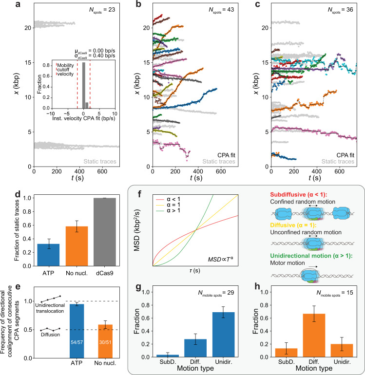
The eukaryotic replicative helicase CMG centrally orchestrates the replisome and leads the way at the front of replication forks. Understanding the motion of CMG on the DNA is therefore key to our understanding of DNA replication. In vivo, CMG is assembled and activated through a cell-cycle-regulated mechanism involving 36 polypeptides that has been reconstituted from purified proteins in ensemble biochemical studies. Conversely, single-molecule studies of CMG motion have thus far relied on pre-formed CMG assembled through an unknown mechanism upon overexpression of individual constituents. Here, we report the activation of CMG fully reconstituted from purified yeast proteins and the quantification of its motion at the single-molecule level. We observe that CMG can move on DNA in two ways: by unidirectional translocation and by diffusion. We demonstrate that CMG preferentially exhibits unidirectional translocation in the presence of ATP, whereas it preferentially exhibits diffusive motion in the absence of ATP. We also demonstrate that nucleotide binding halts diffusive CMG independently of DNA melting. Taken together, our findings support a mechanism by which nucleotide binding allows newly assembled CMG to engage with the DNA within its central channel, halting its diffusion and facilitating the initial DNA melting required to initiate DNA replication.


Conflict of interest statement

The authors declare no competing interests.

Figures

Fig. 1
Fig. 1. Single-molecule imaging of fully reconstituted CMG.
ai Description of hybrid ensemble and single-molecule assay to image fully reconstituted CMG. a-ii Example scan of an optically trapped DNA molecule containing one CMG diffraction-limited spot. b Distribution of numbers of CMG diffraction-limited spots per DNA. c Distribution of numbers of CMG complexes within each diffraction-limited spot. df Distribution of initial positions on the DNA of d all CMG diffraction-limited spots, e diffraction-limited spots containing 2 CMG complexes or f diffraction-limited spots containing 1 CMG complex; the ARS1 origin of replication is indicated by the dashed cyan line. g Example scans separately showing Mcm2-7JF646 diffraction-limited spots (top) and Cdc45LD555 diffraction-limited spots (bottom) on the same DNA molecule. h, i Distributions of initial positions of Mcm2-7JF646 spots and Cdc45LD555 spots on the DNA in the h presence or i absence of DDK. In each condition (with or without DDK), the histograms of Mcm2-7JF646 and Cdc45LD555 initial positions are weighted by the total number of Mcm2-7JF646 spots. j Mean fraction of Mcm2-7JF646 diffraction-limited spots with colocalized Cdc45LD555 diffraction-limited spots in the presence (NMcm2-7 spots = 65) or absence (NMcm2-7 spots = 89) of DDK; error bars show the standard error of proportion. Statistical significance was obtained from a two-sided binomial test (p-value = 2.2 × 10−8).
Fig. 2
Fig. 2. Fully reconstituted CMG exhibits two different motion types.
a Position vs. time plots of dCas9LD555 spots; (inset) distribution of instantaneous velocities coming from the CPA fits of dCas9LD555 spots; red lines show the instantaneous velocity cutoff (5σdCas9) used to separate CMG spots in b and c into static or mobile; CPA fits are not shown for clarity. b, c Position vs. time plots of CMG spots in the b presence of ATP or c absence of nucleotide; CPA fits are plotted in black, static traces are shown in light gray. d Ratio of static CMG traces in the presence of ATP (Nspots = 43), absence of nucleotide (Nspots = 36), and static dCas9 (Nspots = 23) traces; error bars show the standard error of proportion. e Frequency of consecutive CPA segments with the same direction for CMG spots in the presence of ATP (Nmobile spots = 29) or absence of nucleotide (Nmobile spots = 15); inset diagrams illustrate expected segment directions of a unidirectionally moving spot (top) or a diffusive spot (bottom); error bars show the standard error of proportion. f (left panel) Idealized examples of MSD vs. delay time τ plots with an anomalous coefficients α < 1 (red), α = 1 (yellow), and α > 1 (green); (right panel) diagrams illustrating the types of CMG motion corresponding to each of these three cases: constrained diffusion (α << 1), free diffusion (α ≈ 1) or unidirectional motion (α > >1). g, h Fraction of mobile CMG traces classified into different motion types in the g presence of ATP or h absence of nucleotide; error bars show the standard error of proportion.
Fig. 3
Fig. 3. Analysis of CMG motor motion.
a Distribution of absolute instantaneous velocities of unidirectionally moving CMG spots in the presence of ATP; (inset) Distribution of absolute mean velocities of unidirectionally moving CMG spots in the presence of ATP normalized by the length of each trace. b Distribution of processivities of unidirectionally moving CMG spot in the presence of ATP. c Example kymograph of two unidirectionally moving CMG spots that start within the same diffraction-limited spot and split up into two distinct diffraction-limited spots that move along the DNA in opposite directions (Nsplitting events = 2).
Fig. 4
Fig. 4. Nucleotide binding halts CMG diffusion.
a Position vs. time plots of CMG spots in the presence of ATPγS; CPA fits are plotted in black, static traces are shown in light gray and mobile traces are shown in all other colors. b Fraction of static CMG spots in the presence of ATPγS (Nspots = 34); the results from Fig. 2d, are shown as light bars for comparison; error bars show the standard error of proportion. c Model proposed to explain experimental motion results; c-i proposed two populations of CMG present in the ensemble CMG activation reaction; c-ii summary of experimental outcomes in Figs. 2 and 4a, and proposed explanation of their origins. d Fluorescent scan of an SDS-PAGE gel showing the amount of Cdc45LD555 left on linear DNA bound to magnetic beads at one end and containing either a free end or an end capped with a covalently crosslinked methyltransferase. e Densitometry quantification of the experiment shown in d showing the average normalized intensity of three replicates together with their standard deviation. Data points are connected by solid lines to guide the eye. Source data are provided as a Source Data file.

Similar articles

Cited by

References

    1. Bell SP, Labib K. Chromosome duplication in Saccharomyces cerevisiae. Genetics. 2016;203:1027–1067. doi: 10.1534/genetics.115.186452. - DOI - PMC - PubMed
    1. Sánchez, H. et al. DNA replication origins retain mobile licensing proteins. Nat. Commun. 12, 1908 (2021). - PMC - PubMed
    1. Remus D, et al. Concerted loading of Mcm2-7 double hexamers around DNA during DNA replication origin licensing. Cell. 2009;139:719–730. doi: 10.1016/j.cell.2009.10.015. - DOI - PMC - PubMed
    1. Duzdevich D, et al. The dynamics of eukaryotic replication initiation: origin specificity, licensing, and firing at the single-molecule level. Mol. Cell. 2015;58:483–494. doi: 10.1016/j.molcel.2015.03.017. - DOI - PMC - PubMed
    1. Ticau S, Friedman LJ, Ivica NA, Gelles J, Bell SP. Single-molecule studies of origin licensing reveal mechanisms ensuring bidirectional helicase loading. Cell. 2015;161:513–525. doi: 10.1016/j.cell.2015.03.012. - DOI - PMC - PubMed

Publication types