

Development of a Tissue Oxygenation Flow-Based Index Toward Discerning the Healing Status in Diabetic Foot Ulcers
- PMID: 37060195
- PMCID: PMC10654646
- DOI: 10.1089/wound.2022.0170
Development of a Tissue Oxygenation Flow-Based Index Toward Discerning the Healing Status in Diabetic Foot Ulcers
Abstract
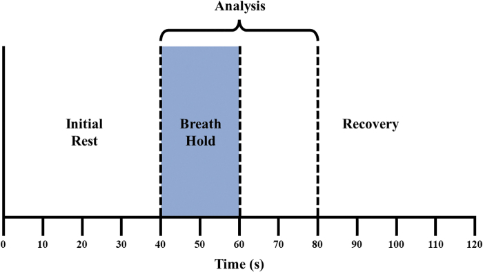
Objective: The objective of this study is to characterize breath-hold (BH)-induced oxygenation changes in diabetic foot ulcers (DFUs) and develop an oxygenation flow index (OFI) to discern nonhealing from healing DFUs. Approach: The imaging approach utilizes an innovative BH stimulus that induces vasoconstriction and measures for altering oxygenation flow in and around the tissues of DFUs and controls. The modified Beer-Lambert law was utilized to calculate hemoglobin-based spatiotemporal oxygenation maps in terms of oxygen saturation. Results: We found controls had synchronous BH-induced oxygenation changes across the dorsal (OFI: 29.0%) and plantar (OFI: 57.6%) aspects of the foot. Nonhealing DFUs, however, had less synchronous BH-induced oxygenation changes (OFI <28%). In addition, two complicated healing DFU cases, or cases with underlying issues or poor long-term healing outcomes, were observed to have OFIs <28%. Innovation: An OFI was developed to differentiate nonhealing DFUs from healing DFUs using a single, noncontact, near-infrared optical scanner for spatiotemporal oxygenation monitoring. The OFI has potential to provide immediate feedback on the microcirculation in DFUs, through hemoglobin-based oxygenation parameters. Conclusion: A preliminary threshold (OFI <28%) could differentiate nonhealing and complicated DFUs from healing DFUs. The overall oxygenation flow pattern was less synchronous (or the OFI value reduced) in the nonwound areas of the feet that were nonhealing. In other words, the reduced OFI value (<28%) in the entire foot, excluding the wound region is a possible indicator that the wound may not heal.
Keywords: breath-hold; diabetic foot ulcers; near infrared spectroscopy; oxygenation flow index; tissue oxygenation.
Conflict of interest statement
There is no conflict of interest with regard to the research in the current article. The corresponding author's university (Florida International University) holds patents on the described NIROS technology and currently filing patents on the proposed innovative techniques related to OFI. The content of this article was expressly written by the authors listed. No ghostwriters were used to write this article.
Figures


Similar articles
-
Breath-Holding as a Stimulus to Assess Peripheral Oxygenation Flow Using Near-Infrared Spectroscopic Imaging.Bioengineering (Basel). 2024 Dec 3;11(12):1221. doi: 10.3390/bioengineering11121221. Bioengineering (Basel). 2024. PMID: 39768039 Free PMC article.
-
Diabetic Foot Ulcer Imaging: An Overview and Future Directions.J Diabetes Sci Technol. 2023 Nov;17(6):1662-1675. doi: 10.1177/19322968231187660. Epub 2023 Aug 18. J Diabetes Sci Technol. 2023. PMID: 37594136 Free PMC article. Review.
-
Breath-Hold Paradigm to Assess Variations in Oxygen Flow in Diabetic Foot Ulcers Using a Noncontact Near-Infrared Optical Scanner.Adv Wound Care (New Rochelle). 2019 Aug 1;8(8):386-402. doi: 10.1089/wound.2018.0922. Epub 2019 Jul 25. Adv Wound Care (New Rochelle). 2019. PMID: 31737422 Free PMC article.
-
The role of CXCL8 in chronic nonhealing diabetic foot ulcers and phenotypic changes in fibroblasts: a molecular perspective.Mol Biol Rep. 2022 Feb;49(2):1565-1572. doi: 10.1007/s11033-022-07144-3. Epub 2022 Jan 19. Mol Biol Rep. 2022. PMID: 35044539 Review.
-
Poorly designed research does not help clarify the role of hyperbaric oxygen in the treatment of chronic diabetic foot ulcers.Diving Hyperb Med. 2016 Sep;46(3):133-134. Diving Hyperb Med. 2016. PMID: 27723012
Cited by
-
Breath-Holding as a Stimulus to Assess Peripheral Oxygenation Flow Using Near-Infrared Spectroscopic Imaging.Bioengineering (Basel). 2024 Dec 3;11(12):1221. doi: 10.3390/bioengineering11121221. Bioengineering (Basel). 2024. PMID: 39768039 Free PMC article.
-
Diabetic Foot Ulcer Imaging: An Overview and Future Directions.J Diabetes Sci Technol. 2023 Nov;17(6):1662-1675. doi: 10.1177/19322968231187660. Epub 2023 Aug 18. J Diabetes Sci Technol. 2023. PMID: 37594136 Free PMC article. Review.
References
-
- Saeedi P, Petersohn I, Salpea P, et al. . Global and regional diabetes prevalence estimates for 2019 and projections for 2030 and 2045: Results from the International Diabetes Federation Diabetes Atlas, 9th edition. Diabetes Res Clin Pract 2019;157:107843; doi: 10.1016/j.diabres.2019.107843 - DOI - PubMed
-
- Anonymous. National Diabetes Statistics Report. Centers for Disease Control and Prevention: Atlanta, Georgia; n.d.
MeSH terms
Substances
LinkOut - more resources
Full Text Sources
Medical
