

Advances in proteomic phenotyping of microglia in neurodegeneration
- PMID: 37060300
- PMCID: PMC10528430
- DOI: 10.1002/pmic.202200183
Advances in proteomic phenotyping of microglia in neurodegeneration
Abstract
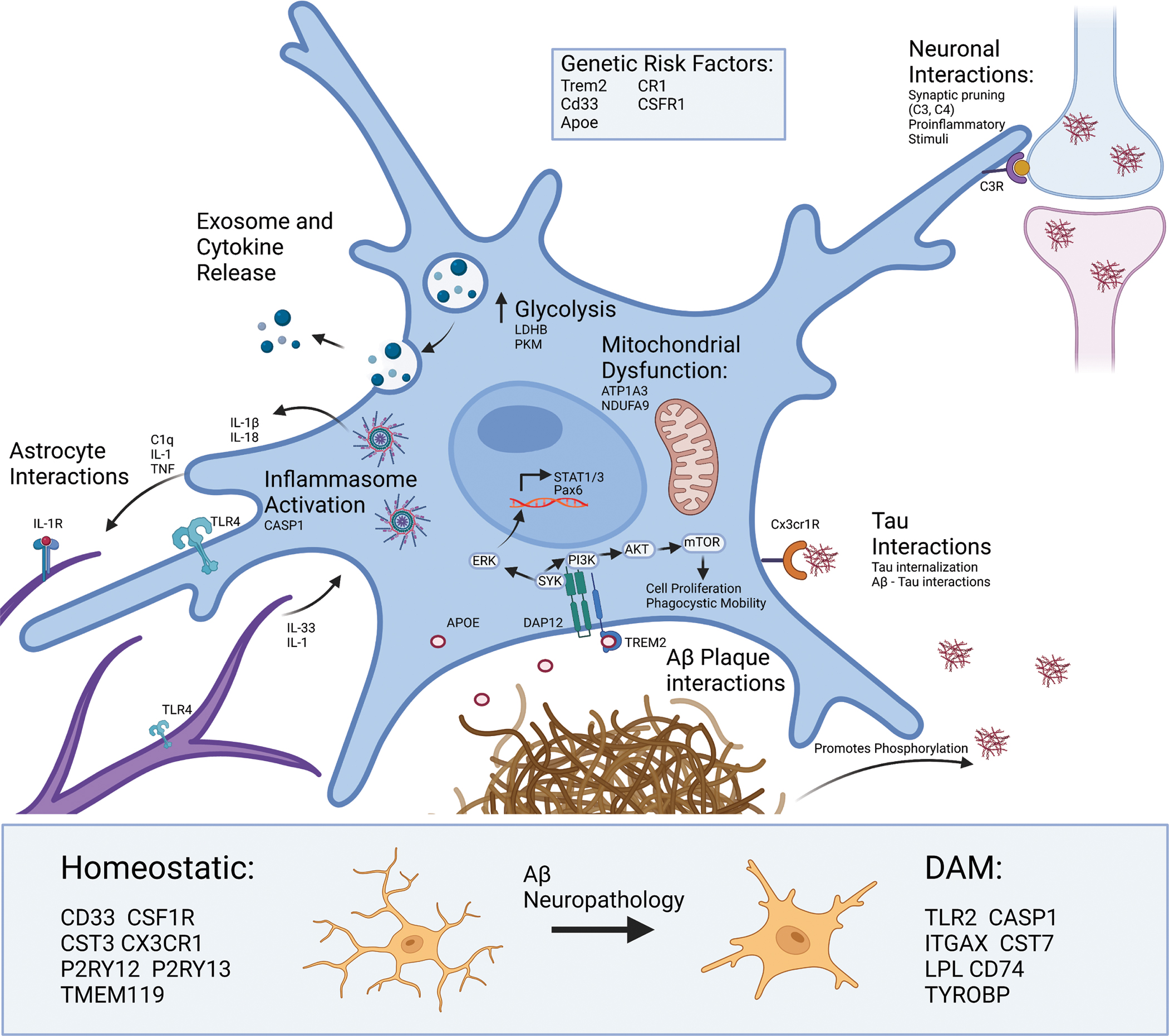
Microglia are dynamic resident immune cells of the central nervous system (CNS) that sense, survey, and respond to changes in their environment. In disease states, microglia transform from homeostatic to diverse molecular phenotypic states that play complex and causal roles in neurologic disease pathogenesis, as evidenced by the identification of microglial genes as genetic risk factors for neurodegenerative disease. While advances in transcriptomic profiling of microglia from the CNS of humans and animal models have provided transformative insights, the transcriptome is only modestly reflective of the proteome. Proteomic profiling of microglia is therefore more likely to provide functionally and therapeutically relevant targets. In this review, we discuss molecular insights gained from transcriptomic studies of microglia in the context of Alzheimer's disease as a prototypic neurodegenerative disease, and highlight existing and emerging approaches for proteomic profiling of microglia derived from in vivo model systems and human brain.
Keywords: inflammation; microglia; neurodegeneration; proteomics; signaling.
© 2023 Wiley-VCH GmbH.
Conflict of interest statement
CONFLICT OF INTEREST STATEMENT
The authors do not have anything to disclose.
Figures
Similar articles
-
Quantitative proteomics of acutely-isolated mouse microglia identifies novel immune Alzheimer's disease-related proteins.Mol Neurodegener. 2018 Jun 28;13(1):34. doi: 10.1186/s13024-018-0266-4. Mol Neurodegener. 2018. PMID: 29954413 Free PMC article.
-
The Functions and Phenotypes of Microglia in Alzheimer's Disease.Cells. 2023 Apr 21;12(8):1207. doi: 10.3390/cells12081207. Cells. 2023. PMID: 37190116 Free PMC article. Review.
-
Microglia, Lifestyle Stress, and Neurodegeneration.Immunity. 2020 Feb 18;52(2):222-240. doi: 10.1016/j.immuni.2019.12.003. Epub 2020 Jan 7. Immunity. 2020. PMID: 31924476 Free PMC article. Review.
-
Microglia in Brain Development, Homeostasis, and Neurodegeneration.Annu Rev Genet. 2019 Dec 3;53:263-288. doi: 10.1146/annurev-genet-112618-043515. Epub 2019 Sep 13. Annu Rev Genet. 2019. PMID: 31518519 Review.
-
Identification and therapeutic modulation of a pro-inflammatory subset of disease-associated-microglia in Alzheimer's disease.Mol Neurodegener. 2018 May 21;13(1):24. doi: 10.1186/s13024-018-0254-8. Mol Neurodegener. 2018. PMID: 29784049 Free PMC article.
Cited by
-
Native-state proteomics of Parvalbumin interneurons identifies unique molecular signatures and vulnerabilities to early Alzheimer's pathology.Nat Commun. 2024 Apr 1;15(1):2823. doi: 10.1038/s41467-024-47028-7. Nat Commun. 2024. PMID: 38561349 Free PMC article.
-
Brain resident microglia in Alzheimer's disease: foe or friends.Inflammopharmacology. 2024 Oct;32(5):2781-2800. doi: 10.1007/s10787-024-01550-8. Epub 2024 Aug 21. Inflammopharmacology. 2024. PMID: 39167311 Review.
References
-
- Penfield W (1932). Cytology & Cellular Pathology of the Nervous System (Vol. 2): PB Hoeber, Incorporated.
Publication types
MeSH terms
Grants and funding
- F31 AG071319/AG/NIA NIH HHS/United States
- RF1AG071587/NH/NIH HHS/United States
- RF1 AG071587/AG/NIA NIH HHS/United States
- R01AG075820/NH/NIH HHS/United States
- F31AG071319/NH/NIH HHS/United States
- F31 NS127530/NS/NINDS NIH HHS/United States
- F31 AG074665/AG/NIA NIH HHS/United States
- R01NS114130/NH/NIH HHS/United States
- F31AG074665/NH/NIH HHS/United States
- R01 AG075820/AG/NIA NIH HHS/United States
- T32 GM135060/GM/NIGMS NIH HHS/United States
- T32 NS096050/NS/NINDS NIH HHS/United States
- R01 NS114130/NS/NINDS NIH HHS/United States
- 1F31NS127530/NH/NIH HHS/United States
LinkOut - more resources
Full Text Sources
Medical
