

Nanomaterial-Based Scaffolds for Tissue Engineering Applications: A Review on Graphene, Carbon Nanotubes and Nanocellulose
- PMID: 37060487
- PMCID: PMC10219911
- DOI: 10.1007/s13770-023-00530-3
Nanomaterial-Based Scaffolds for Tissue Engineering Applications: A Review on Graphene, Carbon Nanotubes and Nanocellulose
Abstract
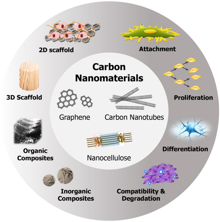
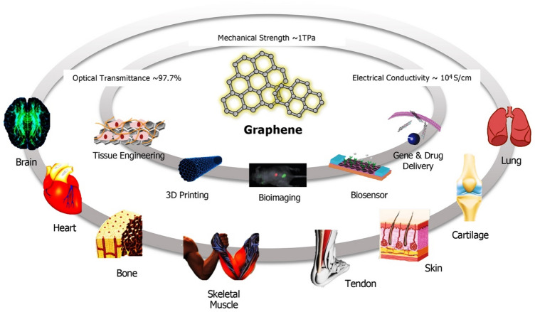
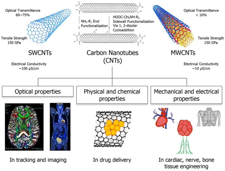
Nanoscale biomaterials have garnered immense interest in the scientific community in the recent decade. This review specifically focuses on the application of three nanomaterials, i.e., graphene and its derivatives (graphene oxide, reduced graphene oxide), carbon nanotubes (CNTs) and nanocellulose (cellulose nanocrystals or CNCs and cellulose nanofibers or CNFs), in regenerating different types of tissues, including skin, cartilage, nerve, muscle and bone. Their excellent inherent (and tunable) physical, chemical, mechanical, electrical, thermal and optical properties make them suitable for a wide range of biomedical applications, including but not limited to diagnostics, therapeutics, biosensing, bioimaging, drug and gene delivery, tissue engineering and regenerative medicine. A state-of-the-art literature review of composite tissue scaffolds fabricated using these nanomaterials is provided, including the unique physicochemical properties and mechanisms that induce cell adhesion, growth, and differentiation into specific tissues. In addition, in vitro and in vivo cytotoxic effects and biodegradation behavior of these nanomaterials are presented. We also discuss challenges and gaps that still exist and need to be addressed in future research before clinical translation of these promising nanomaterials can be realized in a safe, efficacious, and economical manner.
Keywords: Carbon nanotubes; Cellulose nanocrystals; Cellulose nanofibers; Graphene; Regenerative medicine; Tissue engineering.
© 2023. Korean Tissue Engineering and Regenerative Medicine Society.
Conflict of interest statement
All authors declare that they have no conflict of interest.
Figures
Similar articles
-
Graphene nanomaterials as biocompatible and conductive scaffolds for stem cells: impact for tissue engineering and regenerative medicine.J Tissue Eng Regen Med. 2015 Dec;9(12):1321-38. doi: 10.1002/term.1910. Epub 2014 Jun 11. J Tissue Eng Regen Med. 2015. PMID: 24917559 Review.
-
Two- and Three-Dimensional All-Carbon Nanomaterial Assemblies for Tissue Engineering and Regenerative Medicine.Ann Biomed Eng. 2016 Jun;44(6):2020-35. doi: 10.1007/s10439-016-1623-5. Epub 2016 Apr 28. Ann Biomed Eng. 2016. PMID: 27126776 Review.
-
Recent advances in bioactive 1D and 2D carbon nanomaterials for biomedical applications.Nanomedicine. 2018 Oct;14(7):2433-2454. doi: 10.1016/j.nano.2017.03.021. Epub 2017 May 26. Nanomedicine. 2018. PMID: 28552644 Review.
-
A review of carbon nanomaterials/bacterial cellulose composites for nanomedicine applications.Carbohydr Polym. 2024 Jan 1;323:121445. doi: 10.1016/j.carbpol.2023.121445. Epub 2023 Sep 29. Carbohydr Polym. 2024. PMID: 37940307 Review.
-
Versatile Biomaterial Platform Enriched with Graphene Oxide and Carbon Nanotubes for Multiple Tissue Engineering Applications.Int J Mol Sci. 2019 Aug 8;20(16):3868. doi: 10.3390/ijms20163868. Int J Mol Sci. 2019. PMID: 31398874 Free PMC article.
Cited by
-
Correction to "Nanofibrous Microspheres: A Biomimetic Platform for Bone Tissue Regeneration".ACS Appl Bio Mater. 2024 Sep 16;7(9):6325-6331. doi: 10.1021/acsabm.4c01057. Epub 2024 Aug 20. ACS Appl Bio Mater. 2024. PMID: 39162584 Free PMC article. No abstract available.
-
Biomaterial-Based Scaffolds as Carriers of Topical Antimicrobials for Bone Infection Prophylaxis.Pol J Microbiol. 2025 Jun 18;74(2):232-243. doi: 10.33073/pjm-2025-019. eCollection 2025 Jun 1. Pol J Microbiol. 2025. PMID: 40544521 Free PMC article. Review.
-
The potential of copper oxide nanoparticles in nanomedicine: A comprehensive review.Biotechnol Notes. 2024 Jun 5;5:80-99. doi: 10.1016/j.biotno.2024.06.001. eCollection 2024. Biotechnol Notes. 2024. PMID: 39416693 Free PMC article. Review.
-
The Role of Inorganic Nanomaterials in Overcoming Challenges in Colorectal Cancer Diagnosis and Therapy.Pharmaceutics. 2025 Mar 25;17(4):409. doi: 10.3390/pharmaceutics17040409. Pharmaceutics. 2025. PMID: 40284405 Free PMC article. Review.
-
Bridging Gaps in Peripheral Nerves: From Current Strategies to Future Perspectives in Conduit Design.Int J Mol Sci. 2023 May 24;24(11):9170. doi: 10.3390/ijms24119170. Int J Mol Sci. 2023. PMID: 37298122 Free PMC article. Review.
References
-
- Sinha A, Sakon J, Roper DK, Li WJ, Ghosh A, Han HW, et al. Nanoscale particles and multifunctional hybrid soft nanomaterials in bio/nanomedicine. In: Kim JW, Roper DK, Li WJ, editors. Soft matter and biomaterials on the nanoscale. Singapore: World Scientific Publishing; 2020. p. 1–58.
Publication types
MeSH terms
Substances
LinkOut - more resources
Full Text Sources