Review
. 2023 Aug;26(3):313-347.
doi: 10.1007/s10456-023-09876-7. Epub 2023 Apr 15.

Pathological angiogenesis: mechanisms and therapeutic strategies

Affiliations
Review

Pathological angiogenesis: mechanisms and therapeutic strategies

Andrew C Dudley et al. Angiogenesis. 2023 Aug.

Abstract

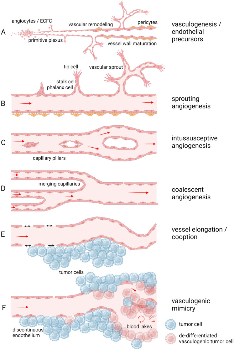
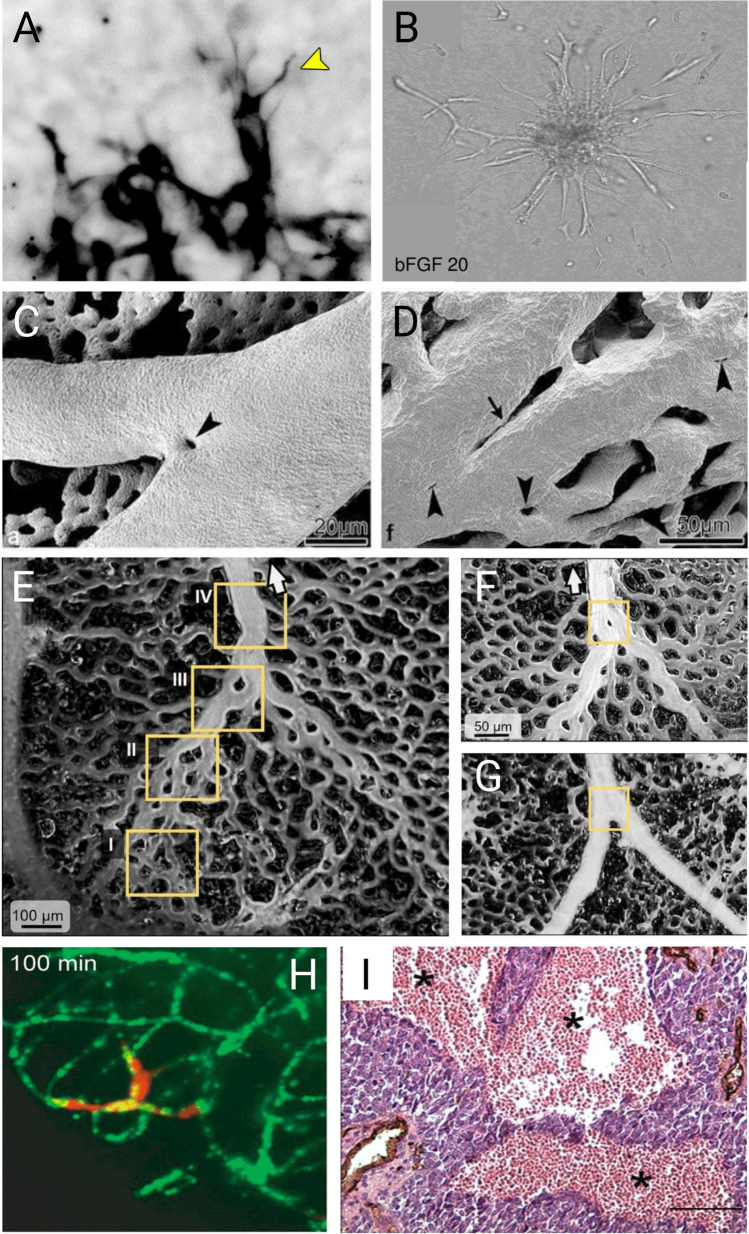
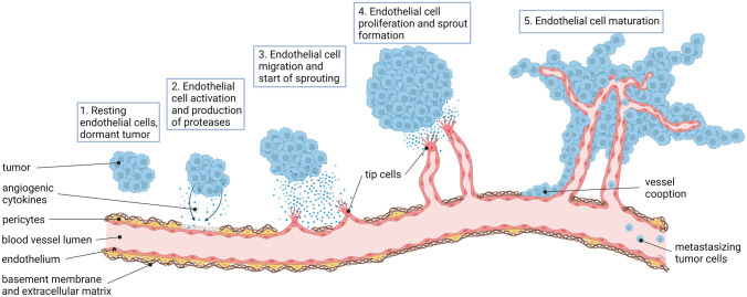
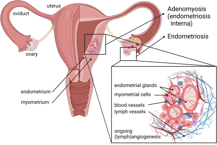
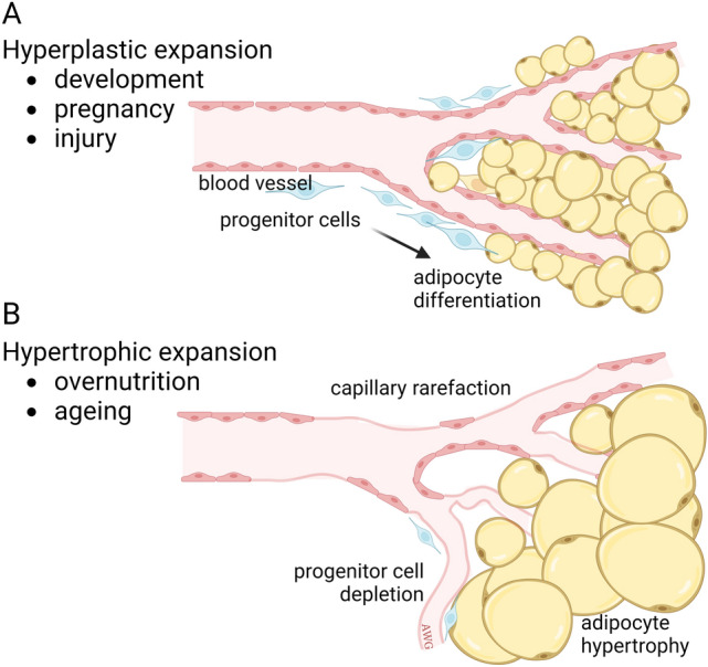
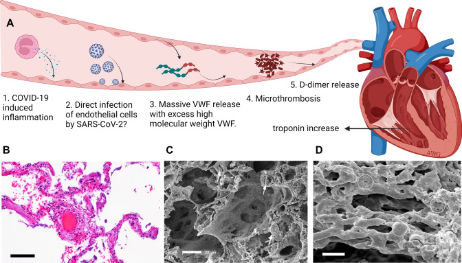
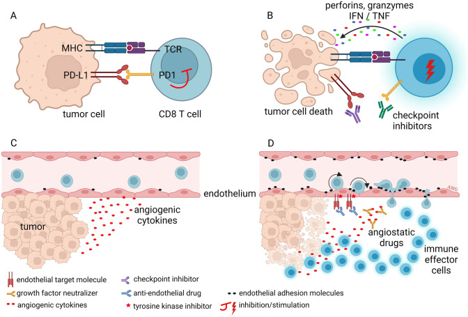
In multicellular organisms, angiogenesis, the formation of new blood vessels from pre-existing ones, is an essential process for growth and development. Different mechanisms such as vasculogenesis, sprouting, intussusceptive, and coalescent angiogenesis, as well as vessel co-option, vasculogenic mimicry and lymphangiogenesis, underlie the formation of new vasculature. In many pathological conditions, such as cancer, atherosclerosis, arthritis, psoriasis, endometriosis, obesity and SARS-CoV-2(COVID-19), developmental angiogenic processes are recapitulated, but are often done so without the normal feedback mechanisms that regulate the ordinary spatial and temporal patterns of blood vessel formation. Thus, pathological angiogenesis presents new challenges yet new opportunities for the design of vascular-directed therapies. Here, we provide an overview of recent insights into blood vessel development and highlight novel therapeutic strategies that promote or inhibit the process of angiogenesis to stabilize, reverse, or even halt disease progression. In our review, we will also explore several additional aspects (the angiogenic switch, hypoxia, angiocrine signals, endothelial plasticity, vessel normalization, and endothelial cell anergy) that operate in parallel to canonical angiogenesis mechanisms and speculate how these processes may also be targeted with anti-angiogenic or vascular-directed therapies.

Keywords: Angiogenesis; Anti-angiogenesis; Endothelial cells; Immunotherapy; Vascular biology; Vascular targeting.


Conflict of interest statement

The authors declare no competing interests.

Figures

Fig. 1
Fig. 1
Different modes of angiogenesis. A The first formation of blood vessels occurs through vasculogenesis and starts early during embryonic development at around E7. The splanchnic layer of the lateral plate mesoderm develops angioblasts, regulated by VEGF production in the endoderm. These angioblasts, the precursors of ECs, become committed to form angioblastic cords that develop into a primitive vascular plexus and subsequently into tubular blood vessels by E9. These early steps of blood vessel development are called vasculogenesis and is followed by different modes of angiogenesis. B Sprouting angiogenesis is the formation of a vascular tree through sprouting ECs from a capillary to form a new capillary bed. In this mode of angiogenesis endothelial tip cells play an important role. C Intussusceptive or splitting angiogenesis is mediated by the formation of an intraluminal pillar by ECs at opposite walls of a capillary. The vessel is longitudinally split into two capillaries, generating two vessels in this way expanding the vascular bed. D Coalescent angiogenesis can be considered the opposite of splitting angiogenesis. Blood vessels coalesce into larger vessels, thereby increasing the efficiency of circulation. E In angiogenic tissues, such as tumors, blood vessels can elongate and become tortuous resulting in an increased vessel density. Blood vessel co-option is the process where cancer cells orchestrate their oxygenation by growing along the well-oxygenized perivascular space. F When cancer cells themselves contribute to vascularization by acquiring EC features, this is called vasculogenic mimicry. This is a rather rare phenomenon, only present in a minor percentage of tumors, but associated with drug resistance and shorter patient survival. Figure is created with BioRender.com and is available on request
Fig. 2
Fig. 2
Modes of angiogenesis imaged. A, B Sprouting angiogenesis. Endothelial tip cells in a mouse lung metastasis (A) and in culture sprouting from a bead in a 3D matrix (B). C, D. Intussusceptive angiogenesis or splitting angiogenesis. ECs form intravascular pillars splitting a vessel into new separate blood vessels. Courtesy of Dr. Djonov, Bern, Switzerland [386]. E, F, G Coalescent angiogenesis. Multiple smaller vessels coalescing into a larger vessel with more efficient blood flow. Courtesy of Drs. Nitzsche and Pries, Berlin, Germany [73]. H Vessel co-option and perivascular migration. The image shows melanoma cells (red) invading along the abluminal surface of the endothelium (green) without evidence of vessel sprouting [79]. I Vasculogenic mimicry. Vascular-like structures formed by cancer cells that upon transdifferentiation can masquerade as ECs. This H&E section shows Ewing sarcoma tissue where vasculogenic mimicry is common and appears as typical “blood lakes”. Blood vessels are stained brown with CD31 antibody [96]
Fig. 3
Fig. 3
Angiogenesis is a hallmark of cancer.1. When a dormant tumor undergoes the angiogenic switch, hypoxia signals induce the production of angiogenic growth factors, such as VEGF, resulting in activation of ECs in nearby blood vessels. 2. Proteases are produced to degrade the ECM around the blood vessels. 3. Migration of ECs is induced and endothelial tip cells guide the EC sprouts into the direction of the growth factor stimulus. 4. Subsequently, proliferation is induced to increase the number of ECs needed for growth of the sprouting neovessels. 5. When vascular sprouts anastomose blood circulation is initiated. The neovasculature is initially immature and leaky, allowing cancer cells to intravasate and metastasize to distant sites. Eventually, EC differentiation, deposition of a functional ECM and attraction of pericytes results in the formation of a mature vasculature. Figure is created with BioRender.com and is available on request
Fig. 4
Fig. 4
Microvasculature in atherosclerosis. A Top images show the progressive development of atherosclerotic plaques in large arteries. Lower images show a progressed but intermediate plaque (B), where blood flow is not blocked and the fibrous cap is strong and stable. At later stages (C) the fibrous cap can become unstable and rupture. This results in the accumulation of thrombocytes, thrombosis, and obstruction of blood flow or even distant embolisms. In healthy conditions, large arteries are vascularized in the outer layers (tunica adventitia) called the vasa vasorum. Plaque formation is initiated by EC dysfunction and accumulation of low density lipoproteins (LDL) in the tunica intima. Expression of EC adhesion molecules recruits monocytes from the blood to form a macrophage infiltrate in the intima of the vessel wall. These become foam cells by accumulating oxidized LDL. Smooth muscle cells migrate into the plaque attracted by immune cell signals as the deposition of a thick fibrous cap develops and microvessels are now attracted by hypoxia signals. With progressing atherosclerosis, the fibrous cap gets thinner and a necrotic core develops. When a plaque ruptures, procoagulant material is exposed, which stimulates thrombus formation. Figure is created with BioRender.com and is available on request
Fig. 5
Fig. 5
Angiogenesis is a feature of endometriosis and adenomyosis. Endometriosis is the presence of endometrium tissue outside the uterus, often resulting from retrograde menstruation. Homing of live endometrial cells and outgrowth into an endometriosis lesion is dependent on angiogenesis. The lesion shown here is present on the ovary, but they can be present anywhere in the peritoneal cavity or even in distant organs. Adenomyosis, or endometriosis interna, is the progressive growth of endometrial glands into the myometrium, supposedly due to microtraumata resulting from the menstrual cycle. Adenomyosis is associated with pain, abnormal bleeding and subfertility [202]. Ectopic endometrium tissue is heavily vascularized, suggesting anti-angiogenic strategies for disease intervention. Figure is created with BioRender.com and is available on request
Fig. 6
Fig. 6
Two mechanisms of adipose tissue expansion. During adipose tissue expansion, cells of the vasculature, adipose progenitor cells, and adipocytes encounter multiple signaling interactions, involving hypoxia, insulin/insulin-like growth factors and vascular guidance cues (apelin/apelin receptor, VEGF, angiopoietins) [212, 387]. A An increase in fat tissue under physiological conditions results from hyperplastic expansion where small adipocytes are generated from multipotent progenitor cells. B Under non-physiological conditions, such as overnutrition and aging, hypertrophic expansion takes place. This is characterized by failing angiogenesis and capillary rarefaction, impairment of progenitor cell proliferation, and hypertrophy of adipocytes. The latter mechanism is strongly associated with metabolic disease risk. Figure adapted from Corvera et al., 2021 [212]. Figure is created with BioRender.com and is available on request
Fig. 7
Fig. 7
SARS-CoV-2 a vascular disease. A Pathophysiology for microthrombosis by SARS-CoV-2 in patients. The figure summarizes the hypothetical steps of the thrombotic sequence from direct or indirect effects of the virus on ECs—this may induce endotheliopathy and a coagulopathy leading to lung obstruction with potential consequences on the right heart ventricle. B H&E staining of a lung of a SARS-CoV-2 patient. Perivascular lymphocytic infiltrate and a microthrombus in an alveolar capillary are seen (bar = 100um). C Scanning electron microscopy demonstrating perivascular and interstitial lymphocytes. Intravascular thrombus was observed in many vessels (white arrows; bar = 200um). D Corrosion casting image showing endothelial injury and endothelialitis. Intraluminal pillars (circles) reflect ongoing intussusceptive angiogenesis (bar = 200um). Figure is adapted from Smadja et al., 2021 [275]. Figure is created with BioRender.com and is available on request
Fig. 8
Fig. 8
Endothelial cell anergy as a vascular immune checkpoint. Immune checkpoint molecules, such as PD-1 and PD-L1, dampen the activity of immune cells. A A tumor cell-specific CD8+ cytotoxic T-cell is prevented from its anti-tumor activity when immune checkpoint molecules are expressed on both cells. B Blocking these molecules by immune checkpoint inhibitory monoclonal antibodies unleashes the anti-tumor activity and cancer cells will be killed. C Angiogenic cancer cells, through secretion of angiogenic growth factors, can downregulate endothelial cell adhesion molecules that can make tumor endothelium unresponsive to proinflammatory cytokines. Such tumor EC anergy results in non-adhesive blood vessels and an immunologically silent tumor microenvironment. Immune suppression based on EC anergy is considered a vascular immune checkpoint. D Inhibition of angiogenesis through growth factor receptor blockade (with tyrosine kinase inhibitors) or neutralization of growth factors (with monoclonal antibodies) overcomes endothelial anergy making cancer cells vulnerable to immune cells. IFN, interferon; PD-1, programmed cell death 1; PD-L1, programmed cell death 1 ligand 1; TCR, T-cell receptor; TNF, tumor necrosis factor. Figure is adapted from Huinen et al., 2021 [158]. Figure is created with BioRender.com and is available on request
Fig. 9
Fig. 9
High endothelial venues (HEVs). A HEVs in secondary lymphoid organs present in the T-cell zone of the lymph node is the location with active extravasation of leukocytes. B HEVs display a cuboidal EC morphology. C An HEV in the inflamed synovium of a rheumatoid arthritis patient. D HEVs in human tonsils, stained for MECA-79 and the HEV nuclear cytokine IL-33 (right). Photomicrographs by courtesy of Drs. Blanchard and Girard, Toulouse, France [338]. Figure is created with BioRender.com and is available on request

Similar articles

Cited by

References

    1. Udan RS, Culver JC, Dickinson ME. Understanding vascular development. Wiley Interdiscip Rev Dev Biol. 2013;2(3):327–346. doi: 10.1002/wdev.91. - DOI - PMC - PubMed
    1. Griffioen AW, Molema G. Angiogenesis: potentials for pharmacologic intervention in the treatment of cancer, cardiovascular diseases, and chronic inflammation. Pharmacol Rev. 2000;52(2):237–268. - PubMed
    1. Pasut A, Becker LM, Cuypers A, Carmeliet P. Endothelial cell plasticity at the single-cell level. Angiogenesis. 2021;24(2):311–326. doi: 10.1007/s10456-021-09797-3. - DOI - PMC - PubMed
    1. Marziano C, Genet G, Hirschi KK. Vascular endothelial cell specification in health and disease. Angiogenesis. 2021;24(2):213–236. doi: 10.1007/s10456-021-09785-7. - DOI - PMC - PubMed
    1. Tzima E, Irani-Tehrani M, Kiosses WB, Dejana E, Schultz DA, Engelhardt B, Cao G, DeLisser H, Schwartz MA. A mechanosensory complex that mediates the endothelial cell response to fluid shear stress. Nature. 2005;437(7057):426–431. doi: 10.1038/nature03952. - DOI - PubMed

Publication types

Substances