. 2023 May 28:562:216178.
doi: 10.1016/j.canlet.2023.216178. Epub 2023 Apr 14.

m6A-modification of cyclin D1 and c-myc IRESs in glioblastoma controls ITAF activity and resistance to mTOR inhibition

Affiliations

m6A-modification of cyclin D1 and c-myc IRESs in glioblastoma controls ITAF activity and resistance to mTOR inhibition

Angelica Benavides-Serrato et al. Cancer Lett. .

Abstract

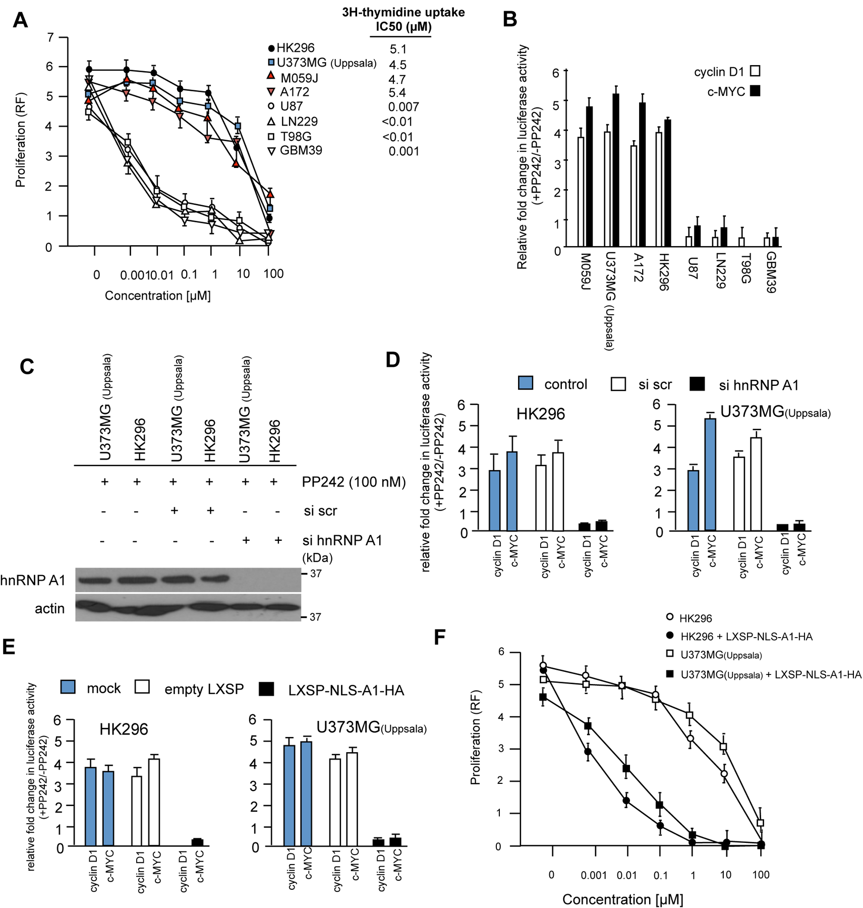
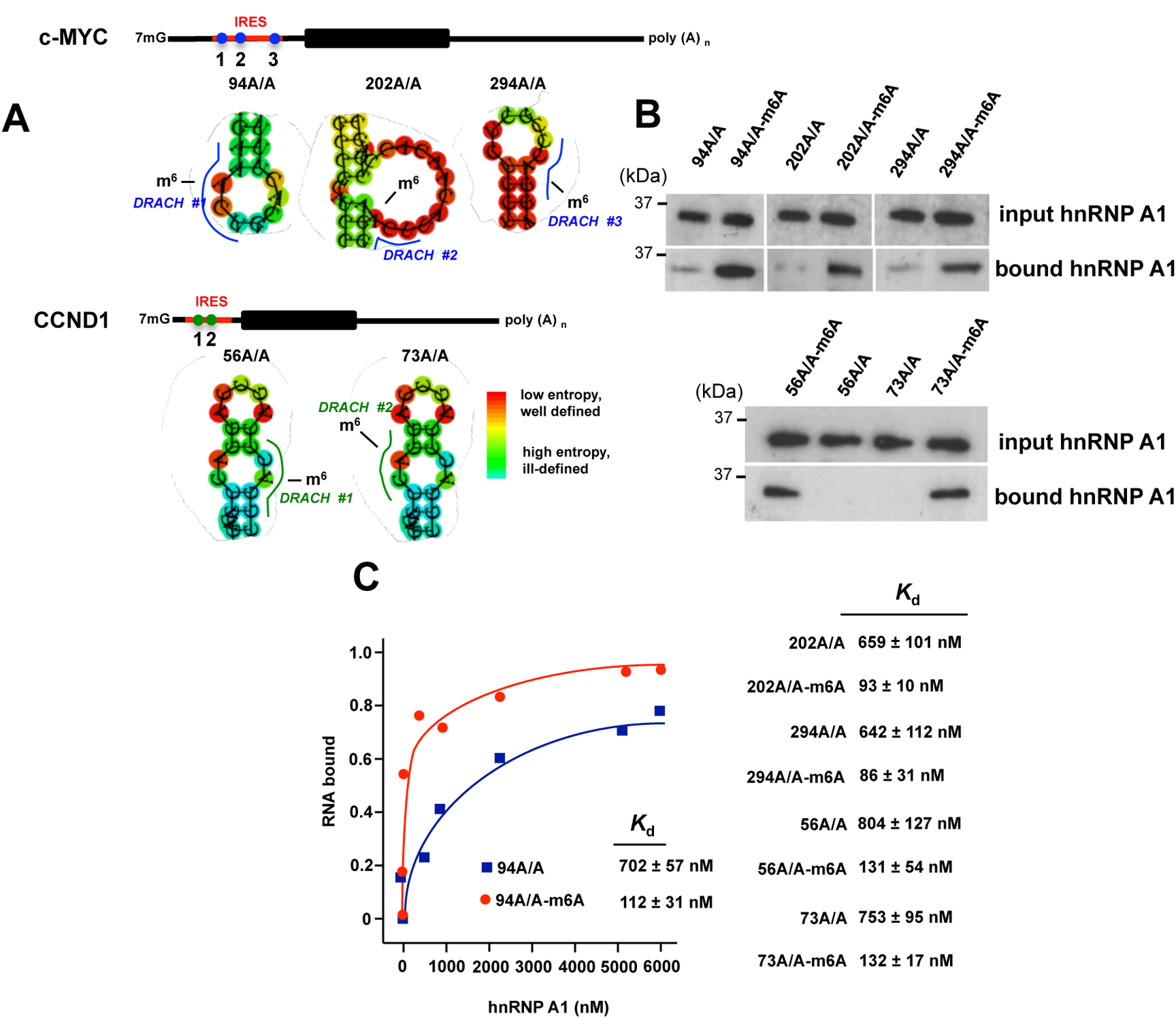
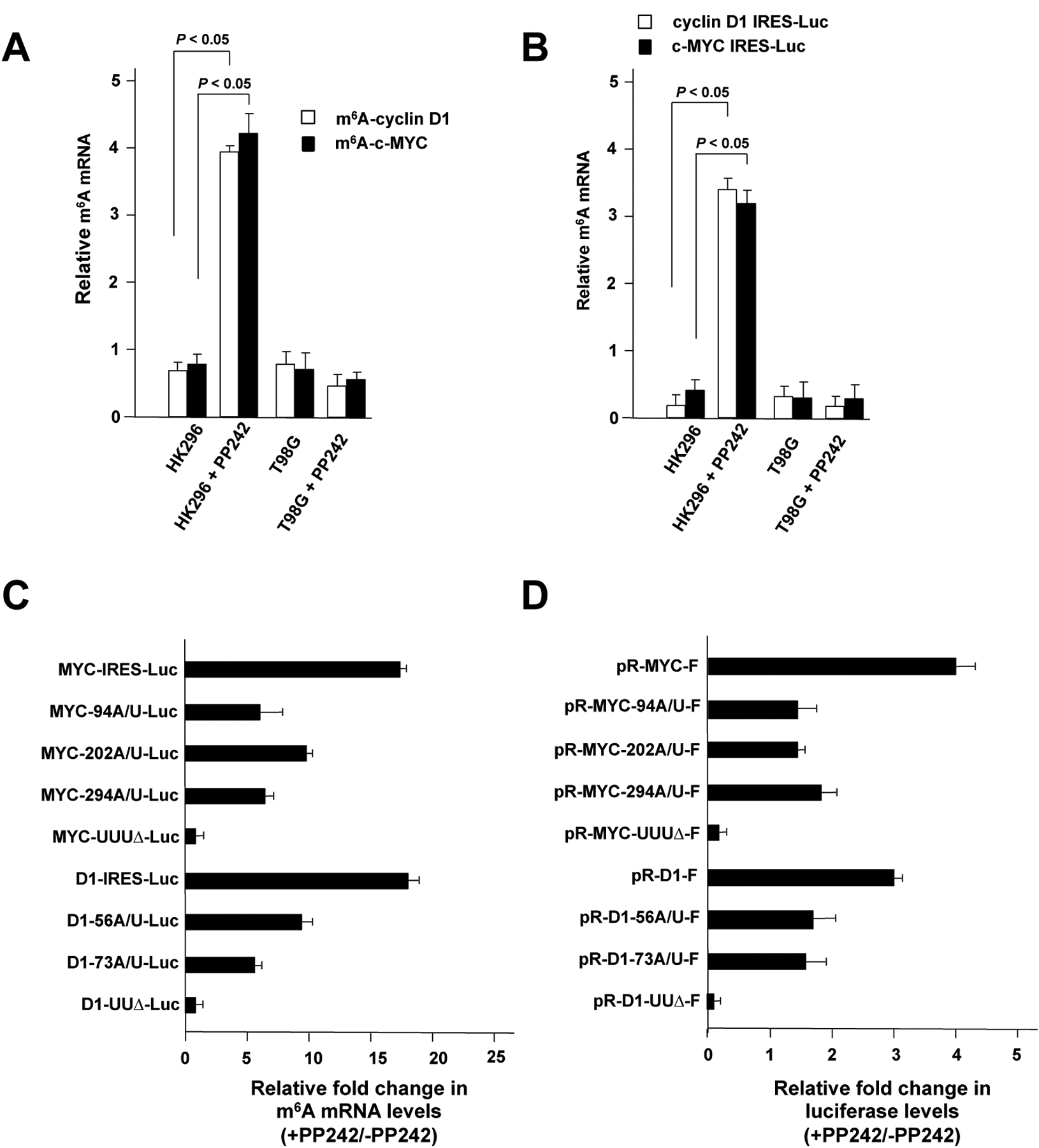
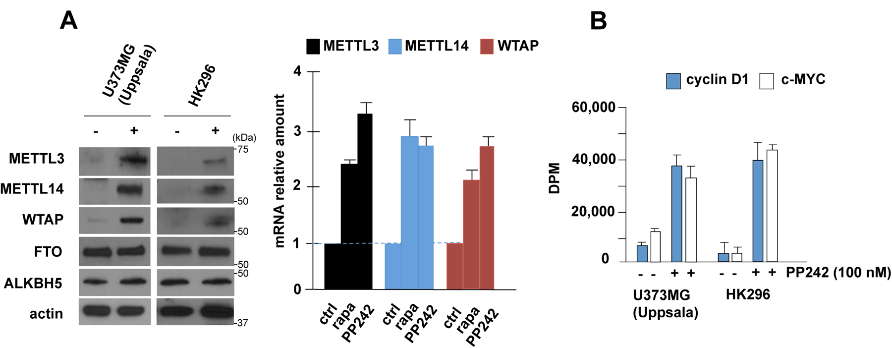
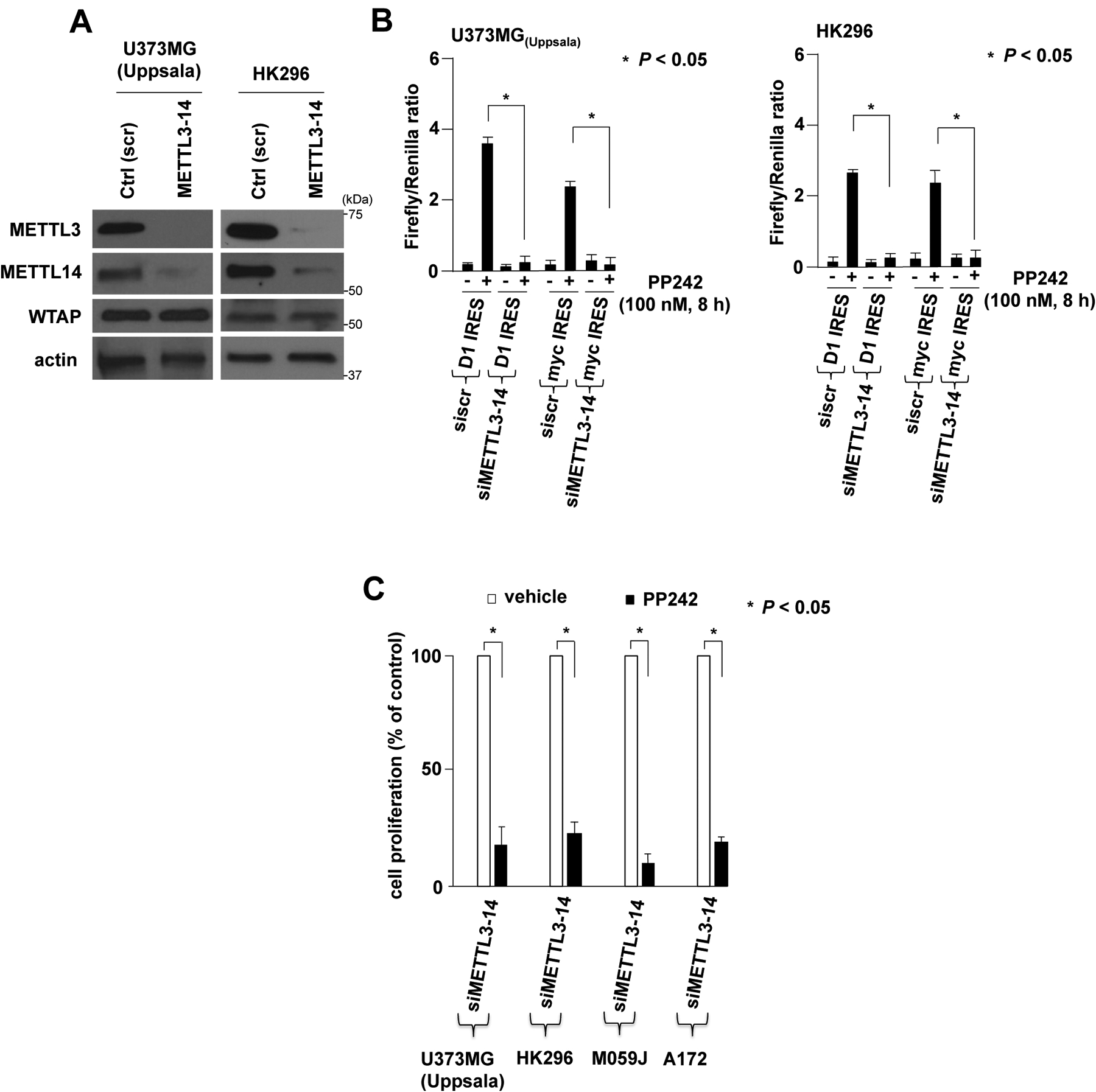
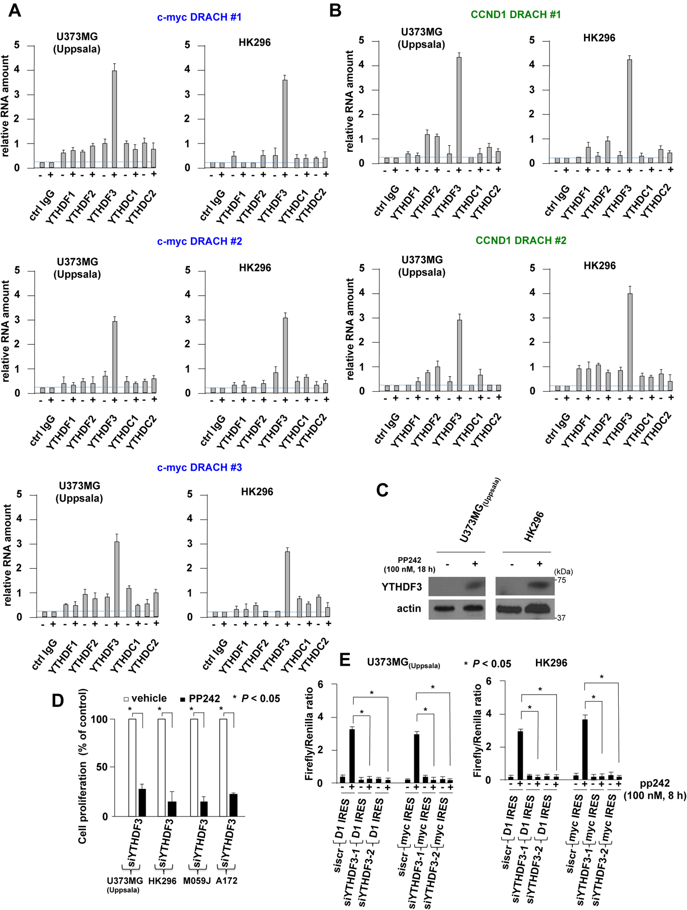
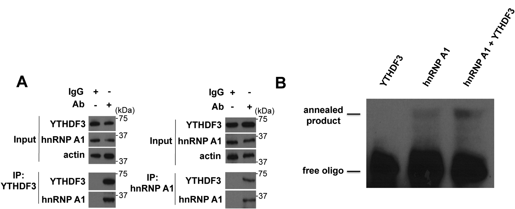
A major mechanism conferring resistance to mTOR inhibitors is activation of a salvage pathway stimulating internal ribosome entry site (IRES)-mediated mRNA translation, driving the synthesis of proteins promoting resistance of glioblastoma (GBM). Previously, we found this pathway is stimulated by the requisite IRES-trans-acting factor (ITAF) hnRNP A1, which itself is subject to phosphorylation and methylation events regulating cyclin D1 and c-myc IRES activity. Here we describe the requirement for m6A-modification of IRES RNAs for efficient translation and resistance to mTOR inhibition. DRACH-motifs within these IRES RNAs upon m6A modification resulted in enhanced IRES activity via increased hnRNP A1-binding following mTOR inhibitor exposure. Inhibitor exposure stimulated the expression of m6A-methylosome components resulting in increased activity in GBM. Silencing of METTL3-14 complexes reduced IRES activity upon inhibitor exposure and sensitized resistant GBM lines. YTHDF3 associates with m6A-modified cyclin D1 or c-myc IRESs, regulating IRES activity, and mTOR inhibitor sensitivity in vitro and in xenograft experiments. YTHDF3 interacted directly with hnRNP A1 and together stimulated hnRNP A1-dependent nucleic acid strand annealing activity. These data demonstrate that m6A-methylation of IRES RNAs regulate GBM responses to this class of inhibitors.

Keywords: Drug resistance; Glioblastoma; IRES; ITAF; N(6)-methyladenosine modification; mTOR inhibitors.


Figures

Fig. 1.
Fig. 1.
Differential sensitivity and dependence on the ITAF hnRNP A1 mediating resistance of GBM lines and short-term primary cultures to PP242. (A) Dose-response curves of the indicated lines following 48 h exposure to PP242. IC50 for resistant cells ranged from 10 – 4.5 μM, while those for sensitive cells ranged from 7 – 12 nM. Proliferation was measured using resazurin. IC50’s as determined via 3H-thymidine uptake assays for each cell line treated with a range of PP242 concentrations are shown to the right of the graph. Mean ± S.D. are shown, n = 3. (B) Relative cyclin D1 and c-myc IRES activities of the indicated GBM lines following 48 h exposure to PP242. Relative fold change in IRES activity derived from dicistronic IRES mRNA reporter assays [20], in which plasmids containing the respective IRES sequences drive IRES dependent translation of Firefly luciferase is shown compared with luciferase activities obtained in the absence of PP242. (C) The ITAF hnRNP A1 is required for PP242-induced IRES activity and drug resistance. siRNA-mediated knockdown of hnRNP A1 in U373MG(Uppsala) and primary HK296 cells. Cells were transiently transfected with siRNAs targeting hnRNP A1 or a nontargeting scrambled (scr) sequence and exposed to PP242 for 24 h and immunoblotted for the indicated proteins. (D) Effects of hnRNP A1 knockdown on PP242-induced IRES activity. U373MG(Uppsala) (right panel) or HK296 (left panel) were transiently transfected with the indicated siRNAs and IRES mRNA reporters and treated with PP242 as shown. Relative fold change in Firefly luciferase activity is shown in the presence of PP242 compared with values obtained in its absence. Mean + S.D. are shown, n = 3. (E) Effects of a dominant-negative shuttling-deficient hnRNP A1 mutant on PP242-induced IRES activity in stably transduced HK296 (left panel) and U373MG(Uppsala) (right panel) cells. The mean and + S.D. are shown for three independent experiments. (F) Ectopic expression of the dominant-negative hnRNP A1 shuttling-deficient mutant confers sensitivity to PP242. The indicated cells stably transduced with the hnRNP A1 mutant was treated with a range of concentrations for 48 h and subsequently proliferation was determined using resazurin. Mean ± S.D. are shown, n = 3.
Fig. 2.
Fig. 2.
Site-specific m6A modification of the cyclin D1 and c-myc IRESs increases hnRNP A1 binding. (A) Schematic of mRNAs, sequences and predicted hairpin structures within the cyclin D1 and c-myc IRESs as indicated. The c-myc IRES harbors three, while the cyclin D1 harbors two well conserved structural motifs containing DRACH (c-myc, blue; cyclin D1 green) sequences as shown. (B) RNA-pull down assays demonstrating that hnRNP A1 preferentially binds m6A methylated cyclin D1 or c-myc IRES RNAs. Biotin conjugated oligonucleotides containing a m6A modification at the appropriate adenosine within the indicated DRACH motifs were used in binding reactions of U373MG(Uppsala) cell lysates and immunoprecipitated using streptavidin coated beads. Immunoprecipitates and input lysates were immunoblotted for hnRNP A1 as indicated. (C) Filter-binding assays showing that m6A modification increases hnRNP A1 binding of the indicated hairpin-loop DRACH containing motifs from the cyclin D1 or c-myc IRESs. The respective dissociation constants (Kd) are indicated in the right panel. Data are mean ± S.D.; n = 3.
Fig. 3.
Fig. 3.
Cyclin D1 and c-myc IRES m6A content is elevated in mTOR inhibitor resistant GBM cells following exposure. (A) Cyclin D1 and c-myc IRES m6A content in HK296 (PP242 resistant) and T98G (PP242 sensitive) cells following exposure to PP242 (100 nM, 4 h). RNAs were extracted from treated cells and immunoprecipitated using α-m6A antibodies. m6A-RNA levels were subsequently determined via qRT-PCR. PCR reactions were performed in quadruplicate and mean + S.D. is shown. P-values are indicated. (B) As in (A) except the indicated cell lines were transfected with either a monocistronic cyclin D1-IRES-luciferase or c-myc-IRES-luciferase mRNA reporter. (C) Relative fold change in m6A-content of the indicated native and mutant IRES RNAs in U373MG(Uppsala) cells in the presence versus absence of PP242 (100 nM, 4h). Mean + S.D. are shown; n = 3. (D) Relative IRES activities of the native cyclin D1 and c-myc IRESs as compared to the indicated hairpin-loop mutants. Relative fold change in IRES activity is shown in the presence versus absence of PP242 treatment in U373MG(Uppsala) cells (100 nM, 4 h). Mean + S.D. are shown; n = 3.
Fig. 4.
Fig. 4.
METTL3–14/WTAP methyltransferase complex expression and activity following mTOR inhibitor exposure in GBM. (A) METTL3, METTL14, WTAP, FTO, ALKBH5 and actin expression following PP242 exposure (100 nM, 18 h) in U373MG(Uppsala) and HK296 cells, left panel. Relative mRNA levels of METTL3, METTL14 and WTAP as determined by qrt-PCR following treatment of U373MG(Uppsala) cells with rapamycin (rapa) (100 nM, 18 h) or PP242 (100 nM, 18 h) as indicated, Mean + S.D., n = 4, right panel. (B) In vitro METTL3–14 activity of the indicated cells following exposure to PP242 as indicated (18 h). c-myc 294 A/A or cyclin D1 73 A/A RNAs were used as substrates in in vitro methylation reactions containing 3H-labeled SAM-e. Mean + S.D., n = 3.
Fig. 5.
Fig. 5.
Silencing the m6A-methylosome blocks IRES activity and leads to mTOR inhibitor sensitivity of resistant GBM tumor cells. (A) RNAi-mediated knockdown of METTL3–14 following transfection of U373MG(Uppsala) or HK296 cells with pooled siRNAs targeting both METTL3 and METTL14 or a non-targeting scrambled control sequence as shown. Lysates were immunoblotted for the indicated proteins. (B) Cyclin D1 or c-myc IRES activity in U373MG(Uppsala) (left panel) or HK296 (right panel) following knockdown of METTL3–14 and treatment with PP242. Activity is displayed as relative Firefly/Renilla luciferase activity from a dicistronic mRNA reporter. Luciferase activity was normalized to the luciferase mRNA level. * P-values are shown. Mean + S.D.; n = 3. (C) Knockdown of METTL3–14 sensitizes PP242-resistant GBM cells. ATP-release assays (Promega CellTiter-Glo® assay) were used to quantify proliferation and displayed as percent of vehicle alone control treatments. * P-values are shown. Mean + S.D. are shown; n = 3.
Fig. 6.
Fig. 6.
YTHDF3 specifically interacts with DRACH RNA motifs in cyclin D1 and c-myc IRESs to regulate IRES activity and mTOR inhibitor sensitivity. (A) Screening of YTH domain family proteins binding to the c-myc DRACH RNA motifs (motifs 1–3, see fig. 2A) in U373MG(Uppsala) or HK296 GBM cells in the absence or presence of PP242 as indicated (100 nM, 18 h). The indicated proteins were immunoprecipitated from lysates and bound RNAs were amplified using primers specific for the hairpin sequences. + S.D., n = 4. (B) as in (A) except binding to the two CCND1 DRACH RNA motifs (motifs 1–2, see fig. 2A) were assessed. (C) Induction of YTHDF3 protein expression following PP242 exposure (100 nM, 18 h) in the indicated lines. (D) siRNA mediated knockdown of YTHDF3 sensitizes PP242 resistant GBM lines to PP242 (100 nM, 48 h). ATP-release assays were performed (Promega CellTiter-Glo® assay) and cell proliferation determined as a percentage of control vehicle treated cells +S.D.; n = 3. (E) siRNA mediated knockdown of YTHDF3 inhibits PP242 induced IRES activity. U373MG(Uppsala) or HK296 cells were transfected with the indicated non-targeting control (scrambled sequence; shscr) or two individual (siYTHDF3–1 or −2) YTHDF3 targeting siRNAs and IRES activity determined from CCND1 or c-MYC dicistronic mRNA reporters in the absence or presence of PP242 as indicated. Luciferase activity was normalized to the luciferase mRNA level. * P-values are shown. Mean + S.D., n = 3.
Fig. 7.
Fig. 7.
hnRNP A1 and YTHDF3 interact and hnRNP A1-induced nucleic acid annealing activity is stimulated by YTHDF3. (A) Extracts from U373MG(Uppsala) cells were immunoprecipitated with nonspecific IgG (control) or antibody against YTHDF3 (left panel) or hnRNP A1 (right panel) and immunoprecipitates immunoblotted for the indicated proteins. Input lysates were probed for the indicated proteins as shown. (B) Recombinant YTHDF3 or hnRNP A1 was purified and analyzed for reannealing activity. 1 pmol of each protein was used in the annealing reactions and the migration positions of the indicated species are displayed.

Similar articles

Cited by

References

    1. Cloughesy TF, Cavenee WK, Mischel PS, Glioblastoma: from molecular pathology to targeted treatment, Annual review of pathology, 9 (2014) 1–25. - PubMed
    1. Omuro A, DeAngelis LM, Glioblastoma and other malignant gliomas: a clinical review, Jama, 310 (2013) 1842–1850. - PubMed
    1. Oronsky B, Reid TR, Oronsky A, Sandhu N, Knox SJ, A Review of Newly Diagnosed Glioblastoma, Frontiers in oncology, 10 (2020) 574012. - PMC - PubMed
    1. Dymova MA, Kuligina EV, Richter VA, Molecular Mechanisms of Drug Resistance in Glioblastoma, International journal of molecular sciences, 22 (2021). - PMC - PubMed
    1. Brennan CW, Verhaak RG, McKenna A, Campos B, Noushmehr H, Salama SR, Zheng S, Chakravarty D, Sanborn JZ, Berman SH, Beroukhim R, Bernard B, Wu CJ, Genovese G, Shmulevich I, Barnholtz-Sloan J, Zou L, Vegesna R, Shukla SA, Ciriello G, Yung WK, Zhang W, Sougnez C, Mikkelsen T, Aldape K, Bigner DD, Van Meir EG, Prados M, Sloan A, Black KL, Eschbacher J, Finocchiaro G, Friedman W, Andrews DW, Guha A, Iacocca M, O’Neill BP, Foltz G, Myers J, Weisenberger DJ, Penny R, Kucherlapati R, Perou CM, Hayes DN, Gibbs R, Marra M, Mills GB, Lander E, Spellman P, Wilson R, Sander C, Weinstein J, Meyerson M, Gabriel S, Laird PW, Haussler D, Getz G, Chin L, The somatic genomic landscape of glioblastoma, Cell, 155 (2013) 462–477. - PMC - PubMed

Publication types

MeSH terms