

Septal annular dilation in chronic ovine functional tricuspid regurgitation
- PMID: 37061178
- PMCID: PMC11088945
- DOI: 10.1016/j.jtcvs.2023.04.003
Septal annular dilation in chronic ovine functional tricuspid regurgitation
Abstract
Introduction: Annular reduction with prosthetic rings represents the current surgical treatment of functional tricuspid regurgitation (FTR). However, alterations of annular geometry and dynamics associated with FTR are not well characterized.
Methods: FTR was induced in 29 adult sheep with either 8 weeks of pulmonary artery banding (PAB, n = 15) or 3 weeks of tachycardia-induced cardiomyopathy (TIC, n = 14). Eight healthy sheep served as controls (CTL). At the terminal procedure, all animals underwent sternotomy, epicardial echocardiography, and implantation of sonomicrometry crystals on the tricuspid annulus (TA) and right ventricular free wall while on cardiopulmonary bypass. Simultaneous hemodynamic, sonomicrometry, and echocardiographic data were acquired after weaning from cardiopulmonary bypass and stabilization. Annular geometry and dynamics were calculated from 3-dimensional crystal coordinates.
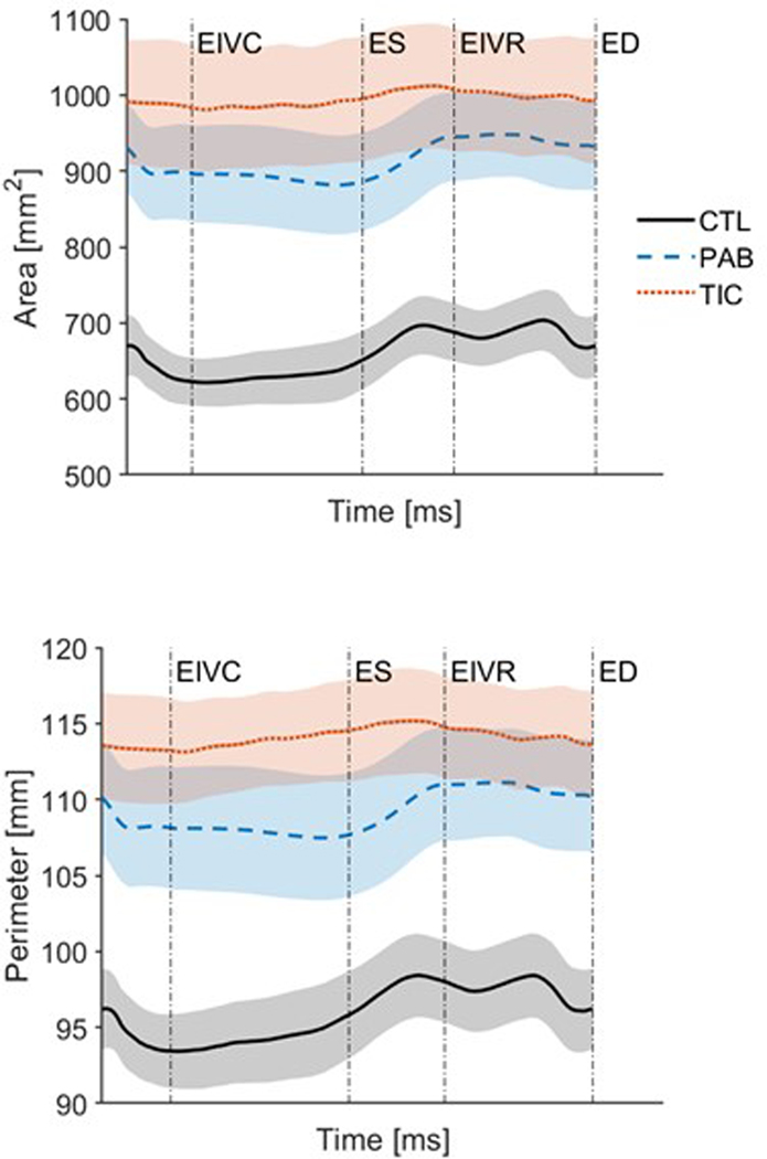
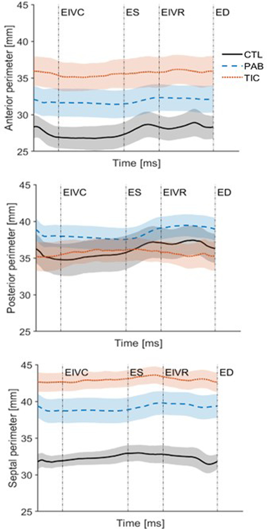
Results: Mean FTR grade (0-4) was 3.2 ± 1.2 and 3.2 ± 0.5 for PAB and TIC, respectively, with both models of FTR associated with similar degree of right ventricular dysfunction (right ventricular fractional area contraction 38 ± 7% and 37 ± 9% for PAB and TIC, respectively). Left ventricular ejection fraction was significantly reduced in TIC versus baseline (33 ± 9%, vs 58 ± 4%, P = .0001). TA area was 651 ± 109 mm2, 881 ± 242 mm2, and 995 ± 232 mm2 for CTL, FTR, and TIC, respectively (P = .006) with TA area contraction of 16.6 ± 4.2%, 11.5 ± 8.0%, and 6.0 ± 4.0%, respectively (P = .003). Septal annulus increased from 33.8 ± 3.1 mm to 39.7 ± 6.4 mm and 43.1 ± 3.2 mm for CTL, PAB, and TIC, respectively (P < .0001).
Conclusions: Ovine FTR was associated with annular dilation and reduced annular area contraction. Significant dilation of septal annulus was observed in both models of FTR. As tricuspid rings do not completely stabilize the septal annulus, continued remodeling may contribute to recurrent FTR after repair.
Keywords: functional tricuspid regurgitation; tricuspid valve; valve repair.
Copyright © 2023 The American Association for Thoracic Surgery. Published by Elsevier Inc. All rights reserved.
Figures


References
-
- Calafiore AM, Foschi M, Kheirallah H, Alsaied MM, Alfonso JJ, Tancredi F, et al. Early failure of tricuspid annuloplasty. Should we repair the tricuspid valve at an earlier stage? The role of right ventricle and tricuspid apparatus. J Card Surg. 2019. Jun;34(6):404–411. doi: 10.1111/jocs.14042. Epub 2019 Apr 8. - DOI - PubMed
-
- Navia JL, Nowicki ER, Blackstone EH, Brozzi NA, Nento DE, Atik FA, et al. Surgical management of secondary tricuspid valve regurgitation: annulus, commissure, or leaflet procedure? J Thorac Cardiovasc Surg. 2010. Jun;139(6):1473–1482.e5. doi: 10.1016/j.jtcvs.2010.02.046. Epub 2010 Apr 14. - DOI - PubMed
Publication types
MeSH terms
Grants and funding
LinkOut - more resources
Full Text Sources
Research Materials
