. 2023 Apr 15;14(1):2168.
doi: 10.1038/s41467-023-37868-0.

Native doublet microtubules from Tetrahymena thermophila reveal the importance of outer junction proteins

Affiliations

Native doublet microtubules from Tetrahymena thermophila reveal the importance of outer junction proteins

Shintaroh Kubo et al. Nat Commun. .

Abstract

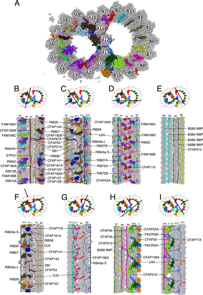
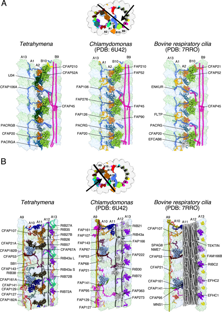
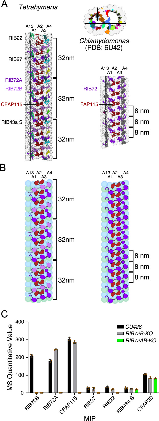
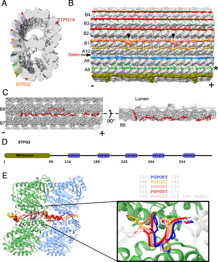
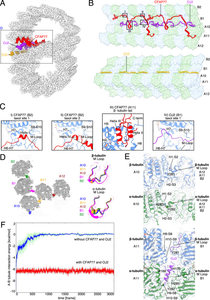
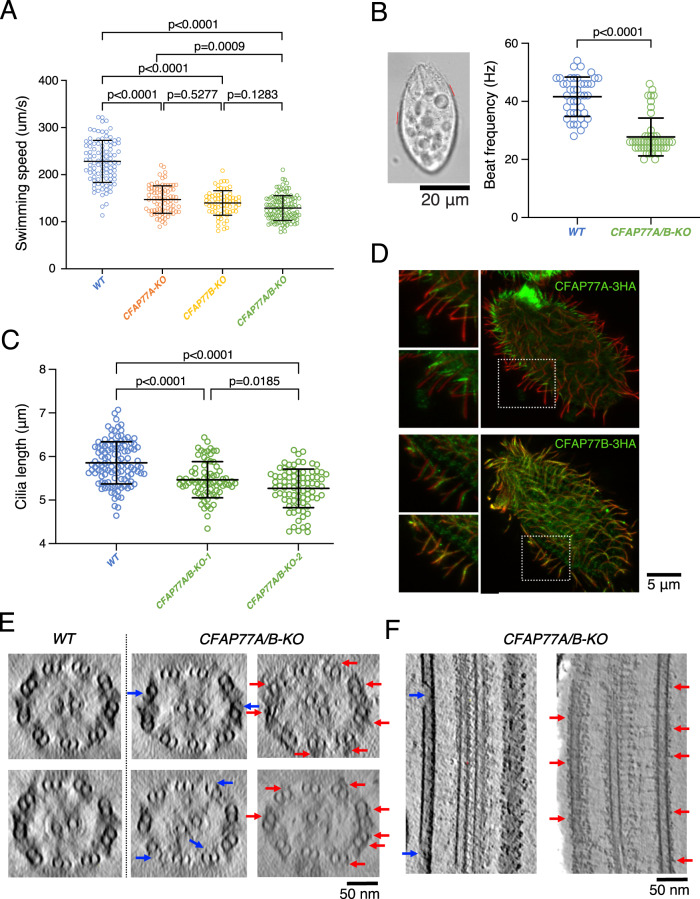
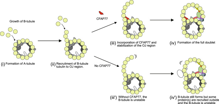
Cilia are ubiquitous eukaryotic organelles responsible for cellular motility and sensory functions. The ciliary axoneme is a microtubule-based cytoskeleton consisting of two central singlets and nine outer doublet microtubules. Cryo-electron microscopy-based studies have revealed a complex network inside the lumen of both tubules composed of microtubule-inner proteins (MIPs). However, the functions of most MIPs remain unknown. Here, we present single-particle cryo-EM-based analyses of the Tetrahymena thermophila native doublet microtubule and identify 42 MIPs. These data shed light on the evolutionarily conserved and diversified roles of MIPs. In addition, we identified MIPs potentially responsible for the assembly and stability of the doublet outer junction. Knockout of the evolutionarily conserved outer junction component CFAP77 moderately diminishes Tetrahymena swimming speed and beat frequency, indicating the important role of CFAP77 and outer junction stability in cilia beating generation and/or regulation.


Conflict of interest statement

The authors declare no competing interests.

Figures

Fig. 1
Fig. 1. The structure of the native DMT from Tetrahymena thermophila.
A A cross-section of the DMT map. Each color denotes an individual MIP. Tubulins are in gray. BI Views of the lumen of the DMT from different angles as indicated by the black arrow. The cutting plane indicated by black lines.
Fig. 2
Fig. 2. Comparison of the DMT structure from Tetrahymena, Chlamydomonas and bovine respiratory cilia.
A The inner junction, note that the architecture is well conserved. B The PF-ribbon region, note many species-specific MIPs.
Fig. 3
Fig. 3. MIPs in Tetrahymena exhibit a 96-nm periodicity.
A TtCFAP115 shows 32-nm repeats leading to the true 96-nm periodicity of the MIPs of Tetrahymena. The FAP115 in Chlamydomonas only repeats with 8-nm periodicity, leading to the normal 48-nm repeat. B The cartoon of Tetrahymena and Chlamydomonas PFs A13-A4 showing the arrangement of FAP115 and RIB72. C Quantitative value of mass spectrometry (normal total spectra value) of WT, RIB72B-KO and RIB72A/B-KO cilia showing that TtCFAP115 is still intact after the knockout of RIB72B. n = 3 biological replicates for WT and each mutant. Data are presented as mean values +/− standard deviation. Values for each replicate are shown in brown circles. Source data are provided as a Source data file.
Fig. 4
Fig. 4. The outer surface filaments on the native DMT.
A, B Cross-sectional (A) and longitudinal (B) views of the outer surface filaments on the native Tetrahymena DMT. The filaments are bound to adjacent pairs of PFs from A8 to B5. The filaments between PFs B2-B3, B3-B4, and B4-B5 appear similar and have a 48-nm periodicity. In contrast, the filaments between PFs A9-A10, A10-B1, and B1-B2 appear to have a 24-nm periodicity. These filaments have a clear head-to-tail periodic arrangement between PFs A9-A10 and B1-B2. The filament between PFs B1B2 has a globular domain (black arrowheads). The density of the filament between PFs A8-A9 is very weak, probably due to partial decoration. (−) and (+) signs indicate the minus and plus ends of the microtubule respectively. C STPG1A is a filamentous protein that is woven between PFs B7-B8, bound to the surface and the lumen. D Schematic representation of the protein topology of the filamentous MIP STPG2. E The PG-rich repeat motifs of STPG2 are structurally similar.
Fig. 5
Fig. 5. CFAP77 stabilizes the outer junction.
A Cross-sectional view of the Tetrahymena outer junction highlighting proteins CFAP77 (red), OJ2 (purple) and OJ3 (yellow). B Architecture of the Tetrahymena outer junction including proteins CFAP77, OJ2 and OJ3. C Helices of CFAP77 occupy taxane-binding pockets of β-tubulin of PF B2. A loop of OJ2 occupies the taxane-binding pocket of β-tubulin of PF B1. The helix-turn-helix motif of CFAP77 is positioned near the C-terminus of β-tubulin of PF A11. D Cross-sectional view of the OJ. M-loops of tubulins are colored according to protofilament: B2 (green); B1 (purple); A10 (blue); A11 (gold); A12 (dark red). To the right, those same M Loops are superimposed. E The canonical (top) and unique (bottom) lateral interactions between tubulin subunits. PFs A11, A12, and B2 are superimposed and adopt the same overall conformation and lateral interactions (top). The lateral interactions between B1 and A11 are particularly unique in Tetrahymena because they involve OJ2 (bottom). F Molecular dynamics simulations of the outer junction with and without CFAP77 and OJ2.
Fig. 6
Fig. 6. Knockout of CFAP77A/B caused mild defects in cilia.
A Knockout of CFAP77A or CFAP77B, or both led up to 40% swimming speed reduction (n = 99 for WT, n = 81 for CFAP77A-KO, n = 70 for CFAP77B-KO, n = 119 for CFAP77A/B-KO). Statistical analyses were done with two-sided Tukey’s multiple comparisons tests. B Tetrahymena cells with marked exemplary positions (red lines) where cilia beat was analyzed in recorded swimming cells. Graphical representation of measurements of cilia beating frequency in WT and CFAP77A/B-KO mutants. (n = 42 cilia from 12 cells for WT, n = 48 cilia from 12 cells for CFAP77A/B-KO). Statistical analyses were done with a two-sided Mann-Whitney comparisons test. C Cilia length measurements of WT and CFAP77A/B-KO mutants. On average cilia length was: WT = 5.85 µm (number of measured cilia, n = 115), CFAP77A/B-KO clone 1 = 5.46 µm (n = 74), CFAP77A/B-KO clone 2 = 5.27 µm (n = 83). Student’s t test WT/KO is 2E−08 and 6,8E−16, respectively. Data are presented as mean values +/− standard deviation in AC. Source data are provided as a Source data file for AC. D Differential localization of CFAP77A and CFAP77B in CFAP77A-3HA and CFAP77B-3HA knock-in mutants. Green—anti-HA; red—poly glycylated tubulin. E The tomographic cross-sections of WT and CFAP77A/B-KO mutants showing occasional damage in the outer junction of CFAP77A/B-KO mutants (blue arrows) and unknown densities near the outer junction region (red arrows). F Longitudinal sections from CFAP77A/B-KO tomogram showing outer junction damage (blue arrows) and unknown densities (red arrows). For (DF), the experiments were done once.
Fig. 7
Fig. 7. A model of the role of CFAP77 on the formation of the B-tubule.
The A-tubule is formed (i), then free tubulins bind to form the hook at the outer junction (OJ) region (ii). With CFAP77, the newly formed B-tubule hook is stabilized (iii), leading to the final DMT formation (iv). Without CFAP77, the B-tubule is not properly stabilized (iii’) and there are additional protein(s) on the outer surface (iv’). The presence of additional protein(s) may or may not be related to elevated levels of tubulin polyglutamylation detected in CFAP77A/B-KO cilia.

Similar articles

Cited by

References

    1. Reiter JF, Leroux MR. Genes and molecular pathways underpinning ciliopathies. Nat. Rev. Mol. Cell Biol. 2017;18:533–547. doi: 10.1038/nrm.2017.60. - DOI - PMC - PubMed
    1. Nicastro D, et al. The molecular architecture of axonemes revealed by cryoelectron tomography. Science. 2006;313:944–948. doi: 10.1126/science.1128618. - DOI - PubMed
    1. Bui KH, Sakakibara H, Movassagh T, Oiwa K, Ishikawa T. Molecular architecture of inner dynein arms in situ in Chlamydomonas reinhardtii flagella. J. Cell Biol. 2008;183:923–932. doi: 10.1083/jcb.200808050. - DOI - PMC - PubMed
    1. Ichikawa M, et al. Tubulin lattice in cilia is in a stressed form regulated by microtubule inner proteins. Proc. Natl Acad. Sci. USA. 2019;116:19930–19938. doi: 10.1073/pnas.1911119116. - DOI - PMC - PubMed
    1. Stepanek L, Pigino G. Microtubule doublets are double-track railways for intraflagellar transport trains. Science. 2016;352:721–724. doi: 10.1126/science.aaf4594. - DOI - PubMed

Publication types

MeSH terms

Substances