

Morphological changes in alveolar bone thickness and height after orthodontic proclination or labial movement combined with autogenous soft tissue grafting: a CBCT evaluation
- PMID: 37061689
- PMCID: PMC10105956
- DOI: 10.1186/s12903-023-02944-w
Morphological changes in alveolar bone thickness and height after orthodontic proclination or labial movement combined with autogenous soft tissue grafting: a CBCT evaluation
Abstract
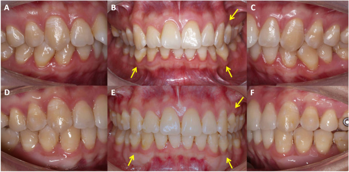
Background: Autogenous soft tissue grafting is indicated in thin gingival biotypes before orthodontic proclination or labial movements to increase the keratinized gingiva and prevent gingival recession. However, its effect on local alveolar bone remodeling is unclear. The aim of this study was to investigate the effects of autogenous soft tissue grafting on local alveolar bone after orthodontic proclination or labial movements.
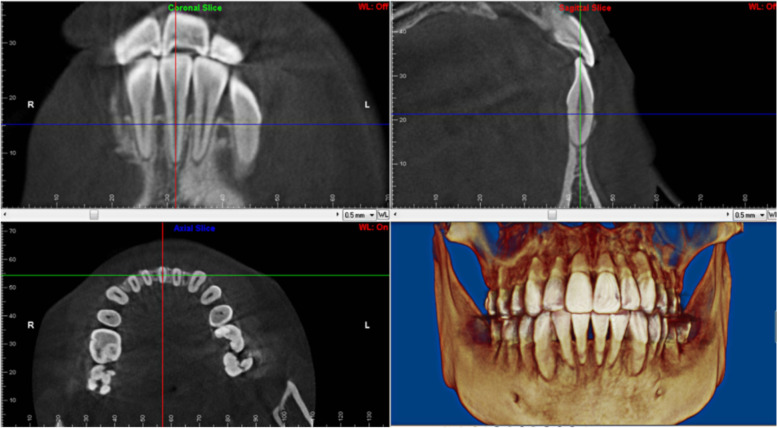
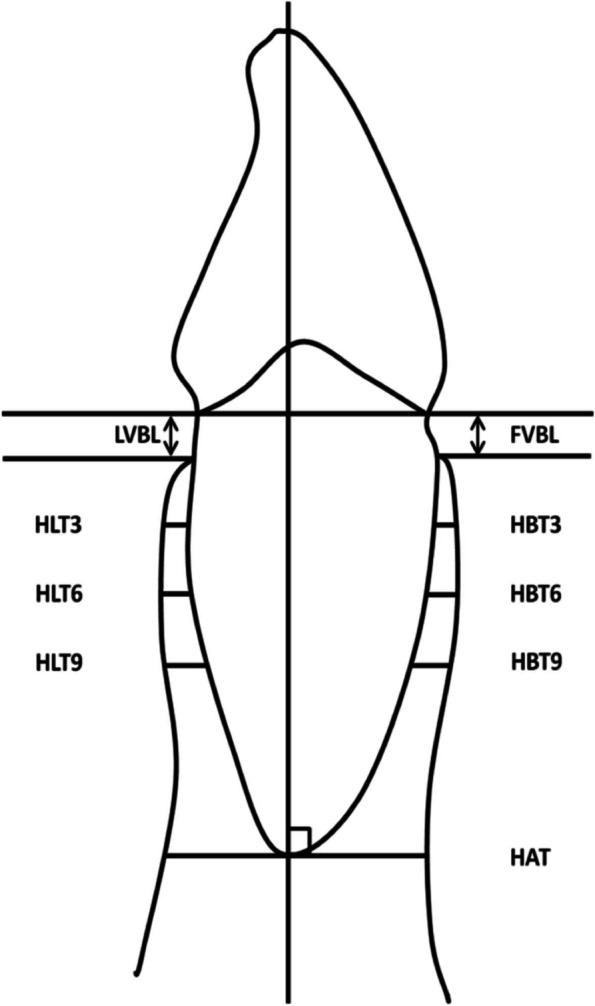
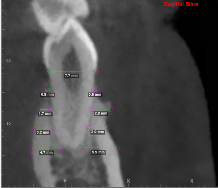
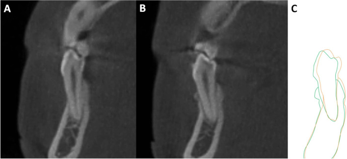
Methods: Sixteen patients with a thin scalloped gingival biotype, narrow keratinized gingiva, or thin cortical bone requiring orthodontic proclination or labial movement of teeth were included. Cone-beam computed tomography (CBCT) images were obtained before grafting and at least 6 months after surgery. Sixty mandibular teeth were included, and the vertical bone level and horizontal labial bone thickness were measured. The results were compared using paired t-tests or Wilcoxon signed-rank test.
Results: The horizontal labial bone thickness increased, especially at 6 mm below the cementoenamel junction (CEJ) in the mandibular central and lateral incisors (P < 0.05). The total alveolar bone area of the canines, first premolars, and second premolars increased at 3, 6, and 9 mm below the CEJ, respectively, and the differences were statistically significant (P < 0.05). Additionally, vertical bone height increased minimally on the labial side, but the differences were not statistically significant (P > 0.05).
Conclusions: New bone regeneration was observed on the labial (pressure) side after autogenous soft tissue grafting, which may represent a mechanism to effectively prevent gingival recession and maintain periodontal health.
Irb approval: All the experimental procedures involving humans in this study were approved by the Medical Ethics Committee of Xiangya Stomatological Hospital, Central South University ( No. 20190048).
Keywords: Autogenous soft tissue grafting; Bone regeneration; Gingival recession; Mucogingival surgery; Orthodontics; Periodontal regeneration.
© 2023. The Author(s).
Conflict of interest statement
The authors declare no competing interests.
Figures
References
-
- Anonymous. Consensus report. Mucogingival therapy. Ann Periodontol. 1996;1(1):702–6. - PubMed
Publication types
MeSH terms
LinkOut - more resources
Full Text Sources
