. 2023 Apr 15;14(1):87.
doi: 10.1186/s13287-023-03302-x.

A beginner's guide on the use of brain organoids for neuroscientists: a systematic review

Affiliations

A beginner's guide on the use of brain organoids for neuroscientists: a systematic review

Lance A Mulder et al. Stem Cell Res Ther. .

Abstract

Background: The first human brain organoid protocol was presented in the beginning of the previous decade, and since then, the field witnessed the development of many new brain region-specific models, and subsequent protocol adaptations and modifications. The vast amount of data available on brain organoid technology may be overwhelming for scientists new to the field and consequently decrease its accessibility. Here, we aimed at providing a practical guide for new researchers in the field by systematically reviewing human brain organoid publications.

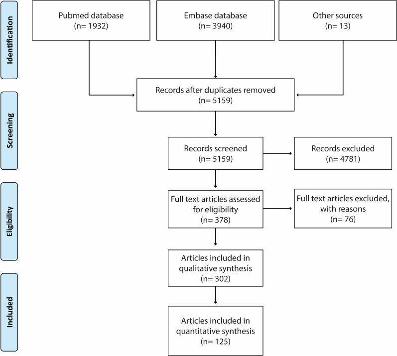
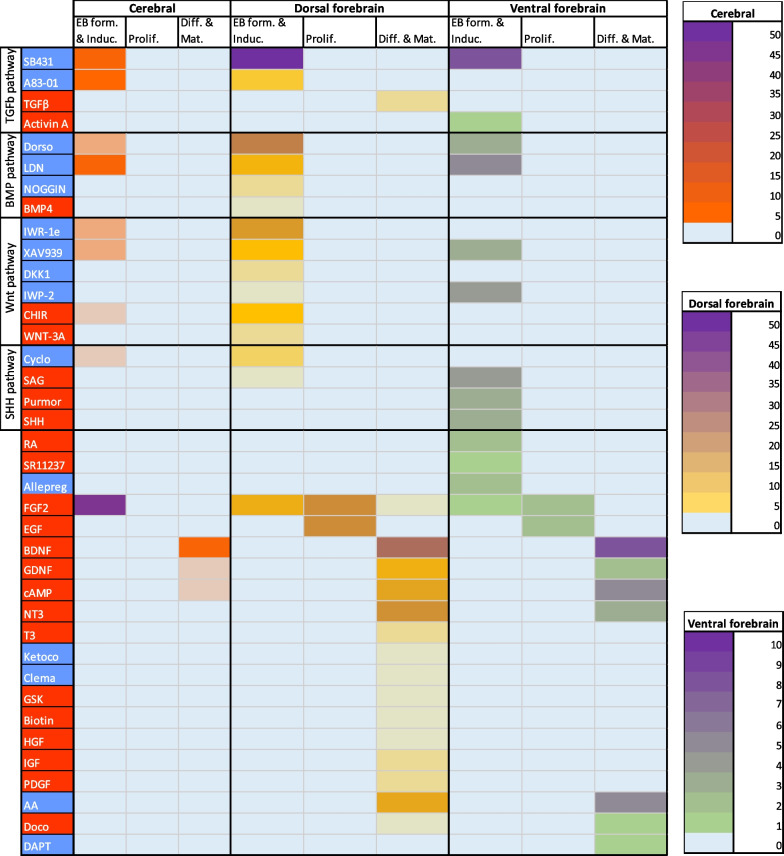
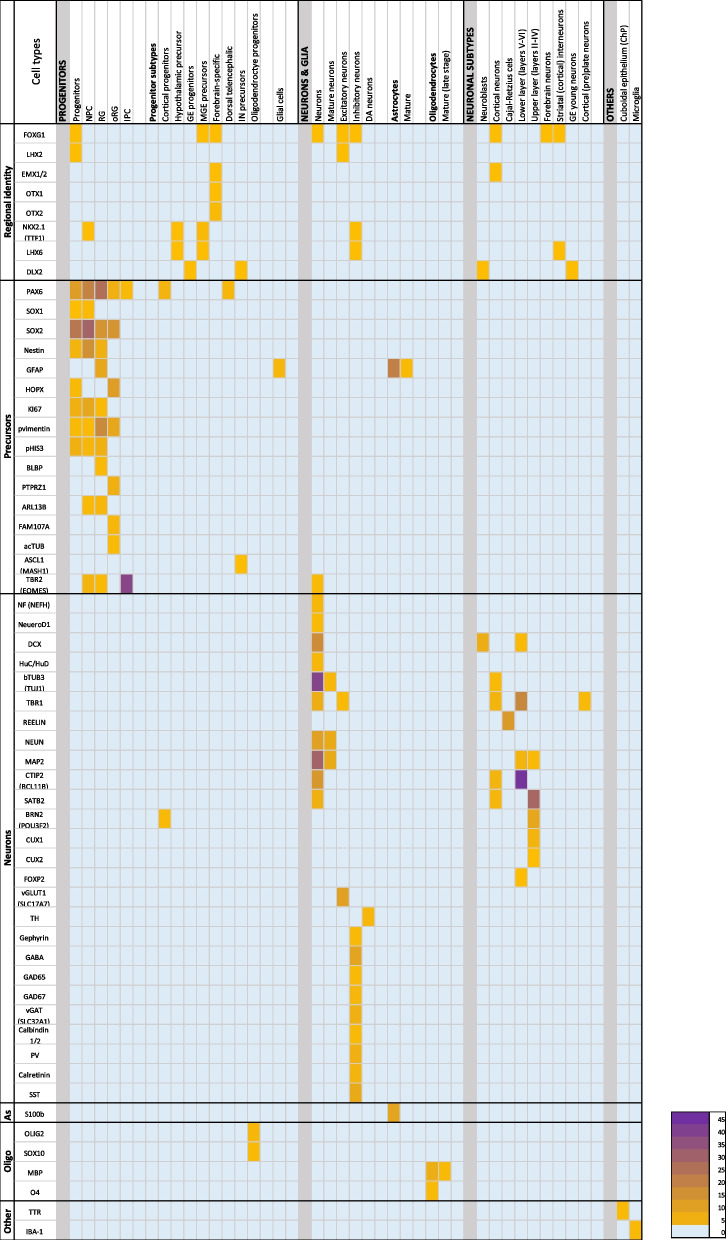
Methods: Articles published between 2010 and 2020 were selected and categorised for brain organoid applications. Those describing neurodevelopmental studies or protocols for novel organoid models were further analysed for culture duration of the brain organoids, protocol comparisons of key aspects of organoid generation, and performed functional characterisation assays. We then summarised the approaches taken for different models and analysed the application of small molecules and growth factors used to achieve organoid regionalisation. Finally, we analysed articles for organoid cell type compositions, the reported time points per cell type, and for immunofluorescence markers used to characterise different cell types.

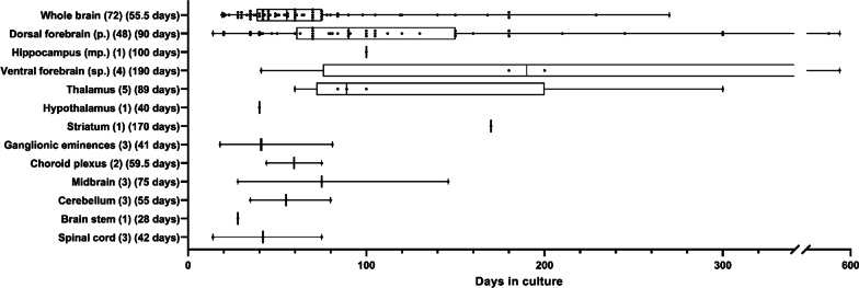
Results: Calcium imaging and patch clamp analysis were the most frequently used neuronal activity assays in brain organoids. Neural activity was shown in all analysed models, yet network activity was age, model, and assay dependent. Induction of dorsal forebrain organoids was primarily achieved through combined (dual) SMAD and Wnt signalling inhibition. Ventral forebrain organoid induction was performed with dual SMAD and Wnt signalling inhibition, together with additional activation of the Shh pathway. Cerebral organoids and dorsal forebrain model presented the most cell types between days 35 and 60. At 84 days, dorsal forebrain organoids contain astrocytes and potentially oligodendrocytes. Immunofluorescence analysis showed cell type-specific application of non-exclusive markers for multiple cell types.

Conclusions: We provide an easily accessible overview of human brain organoid cultures, which may help those working with brain organoids to define their choice of model, culture time, functional assay, differentiation, and characterisation strategies.

Keywords: Cell type characterisation; Human brain organoids; Neurodevelopment; Pluripotent stem cells.


Conflict of interest statement

J.D. & R.S. are employees of uniQure B.V. Other authors (L.M., A.S., K.W. & D.P.) have no competing interest.

Figures

Fig. 1
Fig. 1
Schematic overview of the currently available brain organoid models representing different regions of the human developing central nervous system. The CNS is represented by the forebrain (in dark and light brown), midbrain (green), hindbrain (orange), and spinal Cord (pink). Below each region, the available organoid models are listed with bullet points. Forebrain organoid protocols are subcategorised under telencephalon (dark brown) and diencephalon (light brown) based on the origins of their respective structures. In the forebrain coronary section, the hippocampus is bilaterally depicted with dashed lines in the telencephalon hemispheres. Lining the ventricles is the choroid plexus epithelium (grey line). In the diencephalon, thalamus and hypothalamus are indicated by dashed lines
Fig. 2
Fig. 2
Preferred Reporting Items for Systematic Review and Meta-Analysis for Protocol 2015 article inclusion flow chart
Fig. 3
Fig. 3
Timeline of the first published protocol of different organoid identities. Only first published articles of each brain organoid identity are presented
Fig. 4
Fig. 4
Brain organoid models and their reported days in culture within the neurodevelopmental category are depicted. Box plots depict the 25% and 75% of the individual reports of days in culture, per organoid model. Each report is plotted as a single point. The median days in culture is depicted behind each model for readability. p: pallium, mp: medial pallium, sp: subpallium. The number within brackets depicts the n of articles included per model
Fig. 5
Fig. 5
Small molecule and growth factor used in guided dorsal forebrain and ventral forebrain protocols. Molecules and factors are scored by their uses in EB formation and induction of neuroectoderm, proliferation of the neural tissues, or differentiation and maturation of the organoids. Molecules are grouped by their pathways and determined to exert an inhibitory (blue) or stimulating (red) effect on different pathways. Abbreviations top to bottom, left to right, form: formation, Induc: induction, Prolif: proliferation, Diff: differentiation, Mat: maturation, SB431: SB-431542/3, Activin A: Recominbant Human/ Mouse/ Rat Activin A, Dorso: dorsomorphine, LDN: LDN-193189, IWR-1e: IWR-1(endo), CHIR: CHIR99021, Cyclo: cyclopamine, SAG: smoothened agonist, Purmor: purmorphamine, SHH: Recombinant SHH, RA: retinoic acid, Allepreg: allepregnanolone, Ketoco: ketoconazole, Clema: clemastine, GSK: GSK2656157, HGF: hepatocyte growth factor, IGF: Insulin-like growth factor, PDGF: PDGF-AA, AA: ascorbic acid, Doco: docosahexaenoic acid
Fig. 6
Fig. 6
Cell types and their reported markers. Individual markers are depicted in relation to the cell type that they were reported to characterise. The ‘Regional identity’ column depicts markers not often used for specific cell type characterisations, but more generally used to determine the identity of the organoid model. Strong marker overlap is evident between precursors cells. NPC: neural precursor cells; RG: radial glia; oRG: outer radial glia; IPC: intermediate progenitor cells; GE: ganglionic eminence; MGE: medial ganglionic eminence; IN: interneuron; DA: dopaminergic; ChP: choroid plexus

References

    1. Clancy B, Finlay BL, Darlington RB, Anand KJ. Extrapolating brain development from experimental species to humans. Neurotoxicology. 2007;28(5):931–937. doi: 10.1016/j.neuro.2007.01.014. - DOI - PMC - PubMed
    1. Mellert DJ, Williamson WR, Shirangi TR, Card GM, Truman JW. Genetic and environmental control of neurodevelopmental robustness in drosophila. PLoS ONE. 2016;11(5):e0155957. doi: 10.1371/journal.pone.0155957. - DOI - PMC - PubMed
    1. Hansen DV, Lui JH, Parker PR, Kriegstein AR. Neurogenic radial glia in the outer subventricular zone of human neocortex. Nature. 2010;464(7288):554–561. doi: 10.1038/nature08845. - DOI - PubMed
    1. Lui JH, Hansen DV, Kriegstein AR. Development and evolution of the human neocortex. Cell. 2011;146(1):18–36. doi: 10.1016/j.cell.2011.06.030. - DOI - PMC - PubMed
    1. Ostrem BE, Lui JH, Gertz CC, Kriegstein AR. Control of outer radial glial stem cell mitosis in the human brain. Cell Rep. 2014;8(3):656–664. doi: 10.1016/j.celrep.2014.06.058. - DOI - PMC - PubMed

Publication types

LinkOut - more resources