. 2023 Jul 7;13(7):1572-1591.
doi: 10.1158/2159-8290.CD-22-0620.

Integrative Analysis of a Large Real-World Cohort of Small Cell Lung Cancer Identifies Distinct Genetic Subtypes and Insights into Histologic Transformation

Affiliations

Integrative Analysis of a Large Real-World Cohort of Small Cell Lung Cancer Identifies Distinct Genetic Subtypes and Insights into Histologic Transformation

Smruthy Sivakumar et al. Cancer Discov. .

Abstract

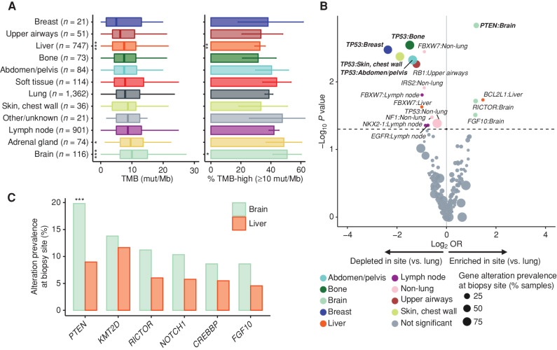
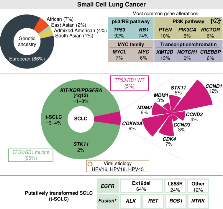
Small cell lung cancer (SCLC) is a recalcitrant neuroendocrine carcinoma with dismal survival outcomes. A major barrier in the field has been the relative paucity of human tumors studied. Here we provide an integrated analysis of 3,600 "real-world" SCLC cases. This large cohort allowed us to identify new recurrent alterations and genetic subtypes, including STK11-mutant tumors (1.7%) and TP53/RB1 wild-type tumors (5.5%), as well as rare cases that were human papillomavirus-positive. In our cohort, gene amplifications on 4q12 are associated with increased overall survival, whereas CCNE1 amplification is associated with decreased overall survival. We also identify more frequent alterations in the PTEN pathway in brain metastases. Finally, profiling cases of SCLC containing oncogenic drivers typically associated with NSCLC demonstrates that SCLC transformation may occur across multiple distinct molecular cohorts of NSCLC. These novel and unsuspected genetic features of SCLC may help personalize treatment approaches for this fatal form of cancer.

Significance: Minimal changes in therapy and survival outcomes have occurred in SCLC for the past four decades. The identification of new genetic subtypes and novel recurrent mutations as well as an improved understanding of the mechanisms of transformation to SCLC from NSCLC may guide the development of personalized therapies for subsets of patients with SCLC. This article is highlighted in the In This Issue feature, p. 1501.


Figures

Figure 1. The genomic landscape of SCLC tumors in a real-world cohort of patients with SCLC. A, Schematic representation of the overall study design in the SCLC cohort comprising 3,600 patients, including 678 cases for which clinical data are available. B, Patterns of the 20 most frequent gene alterations identified in SCLC tumors. Genes are indicated on the left and their alteration frequency on the right. Predicted genomic ancestry, TMB, and microsatellite instability (MSI) status for each patient are overlaid on top. Each sample is color coded based on the type of gene alteration detected. Short variants, such as nonsense, frameshift, and splice alterations, as well as specific rearrangements that result in truncation of the gene product are grouped together as “truncation.” indel, insertions or deletions; MSI-H, high microsatellite instability; MSS, microsatellite stable. C, Prevalence of chromosomal copy-number loss (blue) and gain (red) in SCLC tumors. Notable genes are indicated for some chromosome arms. D, Association between OS and genetic alterations (for genes altered in ≥5 cases) in the clinical cohort of evaluable stage III/IV SCLC (N = 511). Genes identified to be associated with increased survival are shown in green and those identified to be associated with decreased survival are shown in orange (P ≤ 0.05). KDR, PDGFRA, and KIT are all located on 4q12. After FDR-based adjustment, only WHSC1L1 was statistically significant (adjusted P = 0.001). HR, hazard ratio.
Figure 1.
The genomic landscape of SCLC tumors in a real-world cohort of patients with SCLC. A, Schematic representation of the overall study design in the SCLC cohort comprising 3,600 patients, including 678 cases for which clinical data are available. B, Patterns of the 20 most frequent gene alterations identified in SCLC tumors. Genes are indicated on the left and their alteration frequency on the right. Predicted genomic ancestry, TMB, and microsatellite instability (MSI) status for each patient are overlaid on top. Each sample is color coded based on the type of gene alteration detected. Short variants, such as nonsense, frameshift, and splice alterations, as well as specific rearrangements that result in truncation of the gene product are grouped together as “truncation.” indel, insertions or deletions; MSI-H, high microsatellite instability; MSS, microsatellite stable. C, Prevalence of chromosomal copy-number loss (blue) and gain (red) in SCLC tumors. Notable genes are indicated for some chromosome arms. D, Association between OS and genetic alterations (for genes altered in ≥5 cases) in the clinical cohort of evaluable stage III/IV SCLC (N = 511). Genes identified to be associated with increased survival are shown in green and those identified to be associated with decreased survival are shown in orange (P ≤ 0.05). KDR, PDGFRA, and KIT are all located on 4q12. After FDR-based adjustment, only WHSC1L1 was statistically significant (adjusted P = 0.001). HR, hazard ratio.
Figure 2. Biopsy-site-specific patterns of TMB and gene alterations in SCLC. A, Patterns of TMB in SCLC tumors by anatomic location. Shown are box plots of the TMB distribution (left) and the percentage of TMB-high samples (right) at each site. The sites are ordered by their median TMB from lowest (top) to highest (bottom). B, Volcano plot showing patterns of enrichment or depletion of recurrent gene alterations in different tumor sites compared with lung-biopsied SCLC tumors. Gene alterations identified to be statistically different in prevalence between a metastatic site and lung are indicated, with those statistically significant after correcting for multiple hypothesis testing in bold. OR, odds ratio. C, Prevalence and comparison of the most frequently identified gene alterations in brain vs. liver SCLC metastases. (P value thresholds: *, 0.05; **, 0.01; ***, 0.001)
Figure 2.
Biopsy-site-specific patterns of TMB and gene alterations in SCLC. A, Patterns of TMB in SCLC tumors by anatomic location. Shown are box plots of the TMB distribution (left) and the percentage of TMB-high samples (right) at each site. The sites are ordered by their median TMB from lowest (top) to highest (bottom). B, Volcano plot showing patterns of enrichment or depletion of recurrent gene alterations in different tumor sites compared with lung-biopsied SCLC tumors. Gene alterations identified to be statistically different in prevalence between a metastatic site and lung are indicated, with those statistically significant after correcting for multiple hypothesis testing in bold. OR, odds ratio. C, Prevalence and comparison of the most frequently identified gene alterations in brain vs. liver SCLC metastases. (P value thresholds: *, 0.05; **, 0.01; ***, 0.001)
Figure 3. TP53 and RB1 wild-type tumors represent a distinct genetic subtype of SCLC associated with human papilloma virus (HPV) infection. A, Distribution of TMB (mut/Mb) in SCLC tumors wild-type and/or mutant for TP53 and RB1. The TMB in each mutation group was compared against cases identified to be TP53 + RB1 double mutants (P value thresholds: **, 0.01; ***, 0.001). B, Prevalence of TMB-high status (≥10 mut/Mb) in SCLC tumors wild-type and/or mutant for TP53 and RB1. C, Prevalence of gene alterations in SCLC tumors wild-type for TP53 and RB1 compared with TP53- or RB1-mutant SCLC tumors. D, Analysis of the number of mutations representing the tobacco-associated signature (SBS4) in TP53/RB1 wild-type and TP53/RB1-mutant SCLC tumors. E, Comparative prevalence of TP53 and RB1 alterations in patients of EUR and AFR ancestry. F–I, Additional validation of an index case of HPV-positive SCLC detected based on sequencing. The genomic profiling of a liver biopsy showed a TP53 alteration, and a CCNE1 amplification was identified to be wild-type for RB1. Of note, the patient had a prior history of lung adenocarcinoma and prior negative cervical Pap smears. F, Hematoxylin and eosin staining showing the SCLC tumor (200× magnification). G, Immunohistochemistry (IHC) indicating high positivity for p16 (200× magnification). H, IHC indicating retained RB1 (200× magnification). I, PCR-based HPV genotyping from formalin-fixed, paraffin-embedded tumor tissue (two replicates) and a positive control. WT, wild-type.
Figure 3.
TP53 and RB1 wild-type tumors represent a distinct genetic subtype of SCLC associated with human papilloma virus (HPV) infection. A, Distribution of TMB (mut/Mb) in SCLC tumors wild-type and/or mutant for TP53 and RB1. The TMB in each mutation group was compared against cases identified to be TP53 + RB1 double mutants (P value thresholds: **, 0.01; ***, 0.001). B, Prevalence of TMB-high status (≥10 mut/Mb) in SCLC tumors wild-type and/or mutant for TP53 and RB1. C, Prevalence of gene alterations in SCLC tumors wild-type for TP53 and RB1 compared with TP53- or RB1-mutant SCLC tumors. D, Analysis of the number of mutations representing the tobacco-associated signature (SBS4) in TP53/RB1 wild-type and TP53/RB1-mutant SCLC tumors. E, Comparative prevalence of TP53 and RB1 alterations in patients of EUR and AFR ancestry. F–I, Additional validation of an index case of HPV-positive SCLC detected based on sequencing. The genomic profiling of a liver biopsy showed a TP53 alteration, and a CCNE1 amplification was identified to be wild-type for RB1. Of note, the patient had a prior history of lung adenocarcinoma and prior negative cervical Pap smears. F, Hematoxylin and eosin staining showing the SCLC tumor (200× magnification). G, Immunohistochemistry (IHC) indicating high positivity for p16 (200× magnification). H, IHC indicating retained RB1 (200× magnification). I, PCR-based HPV genotyping from formalin-fixed, paraffin-embedded tumor tissue (two replicates) and a positive control. WT, wild-type.
Figure 4. STK11 alterations define a new genetic subtype of SCLC with decreased OS. A, Most frequently detected gene alterations in STK11-mutant SCLC tumors. Gene alterations identified more frequently in STK11-mutant SCLC tumors compared with STK11 wild-type tumors are displayed in mint green, and those identified less frequently in STK11-mutant SCLC tumors compared with STK11 wild-type tumors are in orange. Genes that were similarly prevalent are shown in gray. B, Oncoplot of the most frequent gene alterations identified in STK11-mutant SCLC tumors. Genes are indicated on the left and their alteration frequency on the right. Ancestry and TMB for each case are indicated on top. C, Distribution of TMB (right) and the prevalence of TMB-high status, defined as ≥10 mut/Mb (left), in STK11-mutant and wild-type SCLC tumors. D, Comparative prevalence of STK11 alterations in patients of AFR ancestry compared with other ancestry groups (NA: not available due to low sample counts). E, OS of patients with STK11-mutant and wild-type SCLC tumors. Samples with unknown/ambiguous functional status or reduced quality were excluded from the STK11 wild-type cohort. NA, not available due to low sample counts; WT, wild-type.
Figure 4.
STK11 alterations define a new genetic subtype of SCLC with decreased OS. A, Most frequently detected gene alterations in STK11-mutant SCLC tumors. Gene alterations identified more frequently in STK11-mutant SCLC tumors compared with STK11 wild-type tumors are displayed in mint green, and those identified less frequently in STK11-mutant SCLC tumors compared with STK11 wild-type tumors are in orange. Genes that were similarly prevalent are shown in gray. B, Oncoplot of the most frequent gene alterations identified in STK11-mutant SCLC tumors. Genes are indicated on the left and their alteration frequency on the right. Ancestry and TMB for each case are indicated on top. C, Distribution of TMB (right) and the prevalence of TMB-high status, defined as ≥10 mut/Mb (left), in STK11-mutant and wild-type SCLC tumors. D, Comparative prevalence of STK11 alterations in patients of AFR ancestry compared with other ancestry groups (NA: not available due to low sample counts). E, OS of patients with STK11-mutant and wild-type SCLC tumors. Samples with unknown/ambiguous functional status or reduced quality were excluded from the STK11 wild-type cohort. NA, not available due to low sample counts; WT, wild-type.
Figure 5. Distinct genetic features of SCLC tumors harboring driver oncogenes traditionally associated with NSCLC. A, Oncoplot of the most frequent gene alterations identified in SCLC tumors with kinase-domain mutations in the EGFR gene (n = 107). Genes are indicated on the left, and their alteration frequency is on the right. Ancestry and TMB for each case are indicated on top. B, Distribution of TMB in EGFR-mutant and wild-type SCLC tumors. C, Comparative prevalence of EGFR alterations in patients of EAS ancestry compared with other ancestry groups. D, Analysis of the number of mutations representing APOBEC-associated single-base substitution signatures (SBS2 and SBS13) in EGFR wild-type and mutant SCLC tumors. E, Breakdown of the different EGFR kinase-domain mutations identified in SCLC tumors. F, Oncoplot for SCLC tumors with alterations in known oncogenic drivers of NSCLC, excluding EGFR. Genes identified to be altered in these cases are shown on the left, and their alteration frequency is on the right. TMB and ancestry for each case are overlaid on top. WT, wild-type. G, Genomic and clinicopathologic characteristics of 41 patients with paired NSCLC/SCLC samples. Patients were grouped into four categories based on the patterns of alterations detected: shared alterations with driver (+) in NSCLC and SCLC (orange bar); shared alterations with driver (−) in NSCLC and SCLC (pink bar); shared alterations with driver (+) in NSCLC only and lost/undetected in the matched SCLC (gray bar); and no shared alterations between NSCLC and SCLC (blue bar). Each patient is annotated with the histology, the time between the collection of the NSCLC and SCLC biopsies, the driver alteration (if detected), and TMB. Only genes identified to be altered in ≥2 patients are shown. Bar plots showing the total number of patients with each gene alteration are shown on the right for NSCLC and SCLC samples. LCNEC, large cell neuroendocrine cancer; LUAD, lung adenocarcinoma; nos, not otherwise specified; SCC, squamous cell carcinoma.
Figure 5.
Distinct genetic features of SCLC tumors harboring driver oncogenes traditionally associated with NSCLC. A, Oncoplot of the most frequent gene alterations identified in SCLC tumors with kinase-domain mutations in the EGFR gene (n = 107). Genes are indicated on the left, and their alteration frequency is on the right. Ancestry and TMB for each case are indicated on top. B, Distribution of TMB in EGFR-mutant and wild-type SCLC tumors. C, Comparative prevalence of EGFR alterations in patients of EAS ancestry compared with other ancestry groups. D, Analysis of the number of mutations representing APOBEC-associated single-base substitution signatures (SBS2 and SBS13) in EGFR wild-type and mutant SCLC tumors. E, Breakdown of the different EGFR kinase-domain mutations identified in SCLC tumors. F, Oncoplot for SCLC tumors with alterations in known oncogenic drivers of NSCLC, excluding EGFR. Genes identified to be altered in these cases are shown on the left, and their alteration frequency is on the right. TMB and ancestry for each case are overlaid on top. WT, wild-type. G, Genomic and clinicopathologic characteristics of 41 patients with paired NSCLC/SCLC samples. Patients were grouped into four categories based on the patterns of alterations detected: shared alterations with driver (+) in NSCLC and SCLC (orange bar); shared alterations with driver (−) in NSCLC and SCLC (pink bar); shared alterations with driver (+) in NSCLC only and lost/undetected in the matched SCLC (gray bar); and no shared alterations between NSCLC and SCLC (blue bar). Each patient is annotated with the histology, the time between the collection of the NSCLC and SCLC biopsies, the driver alteration (if detected), and TMB. Only genes identified to be altered in ≥2 patients are shown. Bar plots showing the total number of patients with each gene alteration are shown on the right for NSCLC and SCLC samples. LCNEC, large cell neuroendocrine cancer; LUAD, lung adenocarcinoma; nos, not otherwise specified; SCC, squamous cell carcinoma.
Figure 6. Overview and summary of the main findings from this analysis of 3,600 cases of SCLC. Schematic representation highlighting the main findings from our integrative analysis of 3,600 patients with SCLC with diverse genetic ancestry. WT, wild-type.
Figure 6.
Overview and summary of the main findings from this analysis of 3,600 cases of SCLC. Schematic representation highlighting the main findings from our integrative analysis of 3,600 patients with SCLC with diverse genetic ancestry. WT, wild-type.

Comment in

  • Genetic diversity in small cell lung carcinoma.
    Hayashi T. Hayashi T. Transl Lung Cancer Res. 2024 May 31;13(5):1169-1172. doi: 10.21037/tlcr-24-40. Epub 2024 May 17. Transl Lung Cancer Res. 2024. PMID: 38854933 Free PMC article. No abstract available.

Similar articles

Cited by

References

    1. Rudin CM, Brambilla E, Faivre-Finn C, Sage J. Small-cell lung cancer. Nat Rev Dis Primers 2021;7:3. - PMC - PubMed
    1. Horn L, Mansfield AS, Szczesna A, Havel L, Krzakowski M, Hochmair MJ, et al. . First-line atezolizumab plus chemotherapy in extensive-stage small-cell lung cancer. N Engl J Med 2018;379:2220–9. - PubMed
    1. Paz-Ares L, Dvorkin M, Chen Y, Reinmuth N, Hotta K, Trukhin D, et al. . Durvalumab plus platinum-etoposide versus platinum-etoposide in first-line treatment of extensive-stage small-cell lung cancer (CASPIAN): a randomised, controlled, open-label, phase 3 trial. Lancet 2019;394:1929–39. - PubMed
    1. Howlader N, Forjaz G, Mooradian MJ, Meza R, Kong CY, Cronin KA, et al. . The effect of advances in lung-cancer treatment on population mortality. N Engl J Med 2020;383:640–9. - PMC - PubMed
    1. Zhao X, Kallakury B, Chahine JJ, Hartmann D, Zhang Y, Chen Y, et al. . Surgical resection of SCLC: prognostic factors and the tumor microenvironment. J Thorac Oncol 2019;14:914–23. - PMC - PubMed

Publication types

MeSH terms