Review
. 2023 Sep;33(9):773-787.
doi: 10.1016/j.tcb.2023.02.007. Epub 2023 Apr 14.

Gasdermins gone wild: new roles for GSDMs in regulating cellular homeostasis

Affiliations
Review

Gasdermins gone wild: new roles for GSDMs in regulating cellular homeostasis

Chi G Weindel et al. Trends Cell Biol. 2023 Sep.

Abstract

Since their discovery, members of the gasdermin (GSDM) family of proteins have been firmly established as executors of pyroptosis, with the N-terminal fragment of most GSDMs capable of forming pores in the plasma membrane. More recent findings suggest that some GSDMs can drive additional cell death pathways, such as apoptosis and necroptosis, through mechanisms independent of plasma membrane perforation. There is also emerging evidence that by associating with cellular compartments such as mitochondria, peroxisomes, endosomes, and the nucleus, GSDMs regulate cell death-independent aspects of cellular homeostasis. Here, we review the diversity of GSDM function across several cell types and explore how various cellular stresses can promote relocalization - and thus refunctionalization - of GSDMs.

Keywords: ROS; cardiolipin; cell death–independent gasdermin function; mitochondria; oxidative stress; peroxisomes; phospholipids.


Conflict of interest statement

Declaration of interests The authors declare no competing interests.

Figures

Figure 1
Figure 1. Posttranslational modification sites for the GSDM family of proteins
A. hGSDMA and mGSDMA are cleaved after residues 246 and 247 respectively by the effector protein SpeB [38, 101]. Phosphorylation site at T8 facilitates hGSDMA pore formation [12]. B. hGSDMB is cleaved after D83 by caspase 4 [102] to promote pyroptosis, and after D91 by caspases 3/6/9 during apoptosis [63]. In cytotoxic T cells, granzyme A cleaves GSDMB at residues K229 and K244 [37]. C. hGSDMC is cleaved at D240 by caspases 6/8 to promote pyroptosis [14]. In states of hypoxia, cleavage of hGSDMC by caspase 8 can occur at D365 [103]. D. The canonical pyroptotic cleavage sites for hGSDMD and mGSDMD by caspases 1/4/5/11 are at residues D275 and D276, respectively [25, 27]. mGSDMD is also cleaved by caspase 8 at D276 [104] and at D88 by caspase 3 [32]. mGSDMD is oxidized by ROS at residues C38, C56, C268, C467, and at C192 to enhance pyroptosis [35, 92]. Exocaspase sites L304, L308, V364, and L637 and ubiquitination by SYVN1 at K203 and 204 also enhance hGSDMD activity [105]. Residue F240 is important for dimerization of the hGSDMD p30 fragment [106]. Inhibition of GSDMD occurs through multiple mechanisms: apoptotic caspases 3/7 cleave hGSDMD at Asp87 to prevent pyroptosis [107]. C192 and C39 of mGSDMD, and C191 of hGSDMD are modified by disulfram to block pyroptosis [108]. Itaconation at mGSDMD C77 prevents cleavage and inhibits pyroptosis [109]. hGSDMD can also be cleaved at Q193 by enterovirus 71 to inhibit pyroptosis [110]. Additional autoinhibition points between the N and C terminal of hGSDMD are E15, L192, L290, L373, and L377 [12]. I104 of hGSDMD and I105 of mGSDMD also have inhibitory properties during pyroptosis [13]. Posttranslational modifications to GSDMD in neutrophils include cleavage by cathepsin G at L274 in mGSDMD and by neutrophil elastase at V251 [75, 111]. Neutrophil elastase also cleaves hGSDMD at C268 to promote neutrophil death [75]. E. hGSDME is phosphorylated at T6 to promote pore formation and is cleaved at D270 (mGSDME D271) to promote mitochondrial association and enhance inflammasome activation [34]. F. Oxidation of cysteine residues 328 and 343 are required for pexophagy mediated by mPJVK [18].
Figure 2:
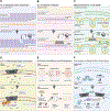
Figure 2:. Shifts in the composition of cellular lipid membranes can impact GSDM binding.
(A) CL in bacterial stress responses. Gram positive bacteria such as S. aureus have high cardiolipin (CL) content in their inner plasma membrane (PM). CL content increases in the context of high osmotic stress, and during infection, with the conversion of phosphatidylglycerol to CL. GSDMs may access/bind to this CL. (B) CL in bacterial virulence. Gram negative bacteria have low CL PM content (<10%). During infection, some species will flip CL to the outer membrane to prevent neutrophil chemotaxis, promote antibiotic resistance, and relocalize effectors to the outer membrane. CL on the outer membrane could serve as a dock for GSDMD association. (C) Mitochondrial CL in cell death. In eukaryotes, CL is localized in the inner mitochondrial membrane (IMM) where it promotes mitochondrial health and IMM structure. During excessive mitochondrial stress caused by apoptotic or pyroptotic stimuli, CL may become localized to the outer mitochondrial membrane (OMM) via CL-flippase, promoting GSDM binding and pore formation. (D) Ca2+ in PM phospholipid conversion. The presence of PI(4,5)P2 on the inner leaflet of the PM enhances GSDM association and promotes dynamic GSDM pores that can open and close. GSDM pore formation results in the influx of Ca2+, which brings PI3K and PLC to the PM where they convert PI(4,5)P2 to DAG and PI(3,4,5)P3– two PL species that GSDMs have reduced affinity for. This limits the dynamic nature of GSDM pores. (E) Endosome remodeling in osteoclastogenesis. Osteoclasts are bone phagocytic cells that reabsorb and break down bone minerals in lysosomes. During late stages of osteoclastogenesis, bone reabsorption is curbed by RANKL-mediated cleavage of caspase-3/8 to generate a non-lytic form of GSDMD(GSDMDp20NT). GSDMDp20NT binds to PI(3)P and prevents its conversion to PI(3,5)P2, a necessary component of late endosomes. (F) Lipid oxidation in GSDMD activation and pyroptosis. The critical antioxidant protein GPX4 maintains lipids in a non-oxidized state. If GPX4 activity is lost, lipid peroxidation occurs resulting in caspase-11 activation, non-canonical inflammasome activation, and GSDMD pore formation. Loss of GPX4 also activates PLCG1, a protein important for PL conversion, to enhance GSDMD activity (although the exact mechanism for this remains uncertain). Figure made with BioRender.
Figure 3:
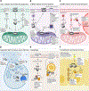
Figure 3:. GSDM organellar associations and their cellular consequences.
(A) CytC release and enhanced apoptosis: Caspases-3/7 cleave GSDME during the extrinsic apoptosis pathway mediated by caspase-8 activation. In addition to associating with the PM, oligomerized GSDMENT is enriched on the mitochondrial network where it promotes the release of cytochrome C (cyt C). Cyt C facilitates both apoptosome mediated apoptotic and pyroptotic cell death and creates a feed forward loop further enhancing caspase-3/7 cleavage and GSDME activation. (B) mtROS release and necroptosis During canonical inflammasome activation in primary mouse macrophages susceptible to ROS stress, GSDMDNT associates with the mitochondria preferentially over the PM. At the mitochondria, GSDMDNT facilitates the release of ROS into the cytosol. Cytosolic ROS activates RIPK3 to both enhance aerobic respiration and mitoROS and trigger the intrinsic necroptotic pathway leading to pMLKL-dependent cell death. (C) mtDNA release and cell cycle disruption. During noncanonical inflammasome activation in endothelial cells, GSDMDNT associates with mitochondria to release mtDNA and activate TBK1 via cGAS cytosolic DNA sensing. TBK1 phosphorylates the transcription factor YAP1 resulting in its degradation, which limits cell proliferation. (D) Neutrophil NET formation and/or NETosis. During NETosis, neutrophils release neutrophil elastase (NE) from azurophilic granules which cleaves GSDMD creating a feed forward loop of NE granule release. GSDMDNT might further enhance NETosis by acting on the nuclear envelope with NE and/or by exerting antimicrobial capabilities in NET extrusions. (E) Autophagy. GSDMs have been shown to impact autophagy in different contexts. High expression of GSDMB is protective in HER2+ cancer cells during lapatinib cancer treatment by promoting LC3 dependent autophagy via RAB7. During sound-induced oxidative stress, auditory hair cells undergo GSDMF-mediated pexophagy via recruitment of LC3 to restrict oxidative damage. (F) Cell adhesion and barrier function In models of intestinal epithelial cell damage linked to inflammatory bowel disease (IBD), methyltrexate (Mtx) treatment upregulates full length GSDMB to the PM where it promotes adhesion, mobility, and epithelial repair via the growth factor PDGF-A and the adhesion protein FAK. Figure made using BioRender.

Similar articles

Cited by

References

    1. Martinon F et al. (2002) The inflammasome: a molecular platform triggering activation of inflammatory caspases and processing of proIL-beta. Mol Cell 10 (2), 417–26. - PubMed
    1. Bjanes E et al. (2021) Genetic targeting of Card19 is linked to disrupted NINJ1 expression, impaired cell lysis, and increased susceptibility to Yersinia infection. PLoS Pathog 17 (10), e1009967. - PMC - PubMed
    1. Kayagaki N et al. (2021) NINJ1 mediates plasma membrane rupture during lytic cell death. Nature 591 (7848), 131–136. - PubMed
    1. Tamura M et al. (2007) Members of a novel gene family, Gsdm, are expressed exclusively in the epithelium of the skin and gastrointestinal tract in a highly tissue-specific manner. Genomics 89 (5), 618–29. - PubMed
    1. Saeki N et al. (2009) Distinctive expression and function of four GSDM family genes (GSDMA-D) in normal and malignant upper gastrointestinal epithelium. Genes Chromosomes Cancer 48 (3), 261–71. - PubMed

Publication types