. 2023 Apr 16;28(1):31.
doi: 10.1186/s11658-023-00443-y.

RPL21 interacts with LAMP3 to promote colorectal cancer invasion and metastasis by regulating focal adhesion formation

Affiliations

RPL21 interacts with LAMP3 to promote colorectal cancer invasion and metastasis by regulating focal adhesion formation

Jiaxian Zhu et al. Cell Mol Biol Lett. .

Abstract

Background: Metastasis is the leading cause of death among patients with colorectal cancer (CRC). Therefore, it is important to explore the molecular mechanisms of metastasis to develop effective therapeutic targets for CRC. In the present study, ribosomal protein L21 (RPL21) was considered as being involved in promoting CRC metastasis, yet the underlying mechanism requires further investigation.

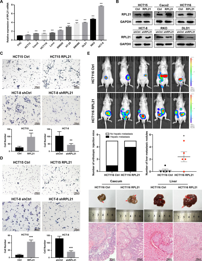
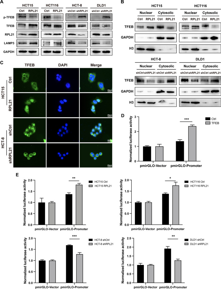
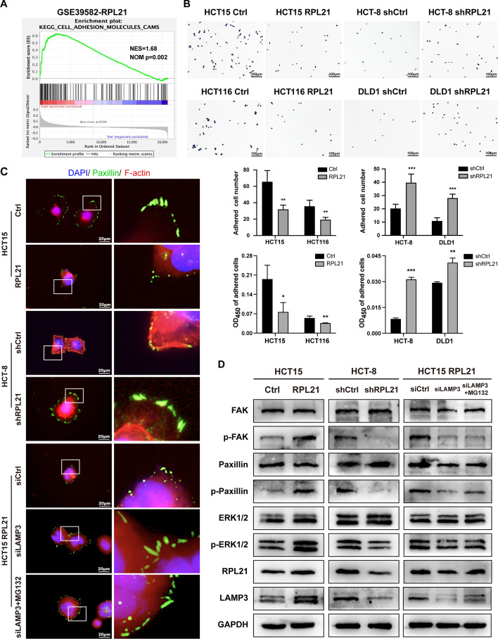
Methods: Immunohistochemistry, western blotting, and quantitative reverse transcription polymerase chain reaction were performed to measure the expression of RPL21 and lysosome-associated membrane protein 3 (LAMP3) in CRC tissues and cells. Wound healing, transwell migration, and invasion assays were performed to study the migration and invasion of cultured CRC cells. An orthotopic CRC mouse model was developed to investigate the metastatic ability of CRC. Transcriptome sequencing was conducted to identify the genes related to RPL21. The dual-luciferase reporter gene assay was performed to determine the transcriptional activity of transcription factor EB (TFEB). The GST/His pull-down assay was performed to investigate the specific binding sites of RPL21 and LAMP3. The cell adhesion assay was performed to determine the adhesion ability of CRC cells. Immunofluorescence staining was performed to observe focal adhesions (FAs).

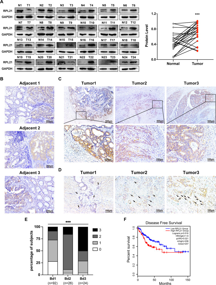
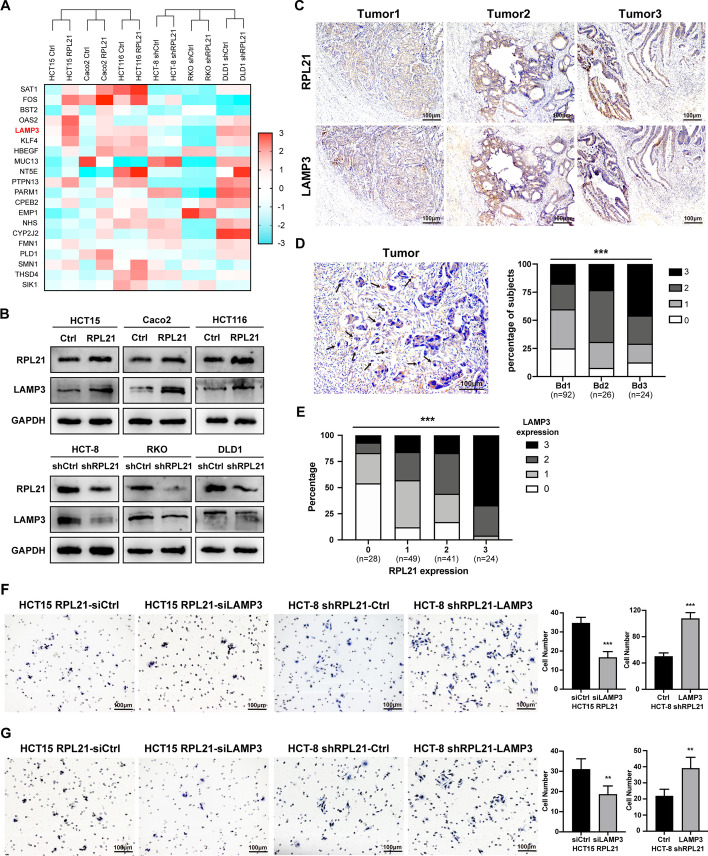
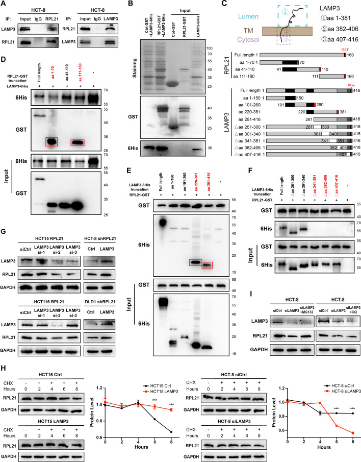
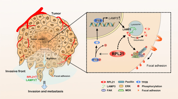
Results: RPL21 was highly expressed in CRC, contributing to tumor invasiveness and poor patient prognosis. Functionally, RPL21 promoted the migration and invasion of CRC cells in vitro and tumor metastasis in vivo. Moreover, LAMP3 was identified as being highly related to RPL21 and was essential in promoting the migration and invasion of CRC cells. Mechanistically, RPL21 activated the transcriptional function of TFEB to upregulate LAMP3 expression. RPL21 directly bound to the aa 341-416 domain of LAMP3 via its aa 1-40 and aa 111-160 segments. The combination of RPL21 and LAMP3 enhanced the stability of the RPL21 protein by suppressing the degradation of the ubiquitin-proteasome system. Furthermore, RPL21 and LAMP3 promoted the formation of immature FAs by activating the FAK/paxillin/ERK signaling pathway.

Conclusions: RPL21 promoted invasion and metastasis by regulating FA formation in a LAMP3-dependent manner during CRC progression. The interaction between RPL21 and LAMP3 may function as a potential therapeutic target against CRC.

Keywords: Colorectal cancer; Focal adhesion; Invasion; LAMP3; Metastasis; Migration; RPL21; TFEB.


Conflict of interest statement

The authors declare that they have no competing interests.

Figures

Fig. 1
Fig. 1
RPL21 is upregulated in CRC tissues, correlating with tumor invasiveness and patient survival rate. A RPL21 protein expression in human CRC tissues (T) and paired adjacent normal tissues (N) was detected by western blotting. The quantification of the protein level was normalized to that of GAPDH. n = 24, ***p < 0.001, paired Student’s t-test. B The representative images of the RPL21 expression in 142 paraffin-embedded human CRC tissues with adjacent normal colorectal tissues (adjacent 1, adjacent 2, and adjacent 3) detected by IHC staining (scale, 200 μm). C The representative images of RPL21 expression in the invasive front of CRC (tumor 1, tumor 2, and tumor 3) were detected by IHC staining (scale, 500 μm, 100 μm). D The representative images of RPL21 expression in CRC tumor buds (tumor 1, tumor 2, and tumor 3) were detected by IHC staining (scale, 100 μm). The arrowheads indicate the tumor buds. E The correlation between the scores of RPL21 expression in tumor buds and the grades of tumor budding. n = 142, ***p < 0.001, Kendall’s Tau test. F Kaplan–Meier survival analysis of CRC patients with high or low expression of RPL21. n = 413
Fig. 2
Fig. 2
RPL21 promotes CRC cell migration, invasion and liver metastasis. A The relative expression of RPL21 mRNA in ten CRC cell lines and FHC was detected by qRT–PCR. Mean ± SD, n = 3, ***p < 0.001, *p < 0.05, Student’s t-test. B The establishment of RPL21 stable overexpression (HCT15, Caco2, and HCT116) and knockdown (HCT-8, RKO, and DLD1) cell lines. RPL21 protein expression was detected by western blotting. C The migration ability of CRC cells was detected by the transwell migration assay (scale, 100 μm). Mean ± SD, n = 5, ***p < 0.001, **p < 0.01, Student’s t-test. D The invasion ability of CRC cells was detected by Matrigel-coated Boyden chamber invasion assay (scale, 100 μm). Mean ± SD, n = 5, ***p < 0.001, Student’s t-test. E In vivo bioluminescence imaging photo and representative morphology images with H&E staining images of tumor formation in mouse cecum and liver metastasis (scale, 100 μm), using the orthotopic CRC mouse model injected with HCT116 Ctrl or HCT116 RPL21 cells. The red arrowheads indicate the tumors in situ or the liver metastatic nodules. The number of liver metastatic nodules in each mouse was counted under the microscope. Mean ± SD, n = 5, *p < 0.05, Student’s t-test
Fig. 3
Fig. 3
LAMP3 is significantly correlated with RPL21 and promotes the migration and invasion of CRC. A Heatmap of the top 20 differentially expressed genes in the transcriptome sequencing of RPL21 stable overexpression and knockdown cells with their control cells. B LAMP3 protein expression in RPL21 stable overexpression and knockdown cells detected by western blotting. C The representative images of RPL21 and LAMP3 expression in 142 paraffin-embedded human CRC tissues (tumor 1, tumor 2, and tumor 3) detected by IHC staining (scale, 100 μm). D The representative image of LAMP3 expression in CRC tumor buds was detected by IHC staining (scale, 100 μm). The arrowheads indicate the tumor buds. The correlation between the scores of LAMP3 expression in tumor buds and the grades of tumor budding was analyzed. n = 142, ***p < 0.001, Kendall’s Tau test. E The correlation between the scores of RPL21 and LAMP3 expression in tumor buds was analyzed. n = 142, ***p < 0.001, Kendall’s Tau test. F The migration ability of the indicated CRC cells was detected by the transwell migration assay (scale, 100 μm). Mean ± SD, n = 5, ***p < 0.001, Student’s t-test. G The invasion ability of indicated CRC cells was detected by Matrigel-coated Boyden chamber invasion assay (scale, 100 μm). Mean ± SD, n = 5, **p < 0.01, Student’s t-test
Fig. 4
Fig. 4
RPL21 upregulates LAMP3 expression by activating TFEB. A TFEB, p-TFEB (Ser211), RPL21, and LAMP3 protein expression in the indicated CRC cells was detected by western blotting. B The TFEB protein expression in cytosolic and nuclear of the indicated CRC cells was detected by western blotting. The quantification of cytosolic TFEB was normalized to GAPDH, the quantification of nuclear TFEB was normalized to H3. C The localization of TFEB (green) in the indicated CRC cells was observed by IF staining (scale, 20 μm). D The transcription of LAMP3 by TFEB was verified by dual-luciferase reporter gene assay in HEK293T cells. Mean ± SD, n = 3, ***p < 0.001, Student’s t-test. E The transcription of LAMP3 in the indicated CRC cells was verified by dual-luciferase reporter gene assay. Mean ± SD, n = 3, ***p < 0.001, **p < 0.01, *p < 0.05, Student’s t-test
Fig. 5
Fig. 5
LAMP3 directly interacted with RPL21 and enhanced the stability of the RPL21 protein. A RPL21 and LAMP3 were immunoprecipitated in HCT-8 cells detected by western blotting. B Direct binding of RPL21-GST and LAMP3-6His using the His pull-down assay. The pull-down bands were detected by Coomassie brilliant blue staining and western blotting. C Topology of LAMP3, including the luminal segment (aa 1–381), the transmembrane segment (aa 382–406), and the cytoplasmic segment (aa 407–416). Diagrammatic representation of RPL21 and LAMP3 with their truncated/deleted mutation forms. DF HEK293T cells were transfected with the indicated constructs and immunoprecipitated with anti-6His (D) or anti-GST (E, F). The red boxes represent the pull-down bands detected by western blotting. G LAMP3 and RPL21 protein expression in the indicated cells was detected by western blotting. H The degradation of the RPL21 protein in HCT15 Ctrl, HCT15 LAMP3, HCT-8 siCtrl, and HCT-8 siLAMP3 cells with CHX treatment (50 μg/mL) was detected by western blotting. The quantification of the protein level was normalized to that of GAPDH. Mean ± SD, n = 3, ***p < 0.001, Student’s t-test. I LAMP3 and RPL21 protein expression in HCT-8 siCtrl, HCT-8 siLAMP3, and HCT-8 siLAMP3 cells treated with MG132 (10 μM, 4 h) or CQ (50 μM, 2 h) was detected by western blotting
Fig. 6
Fig. 6
RPL21–LAMP3 regulates FA formation and promotes cell spreading. A KEGG enrichment analysis of the RPL21 expression in the CRC database GSE39582. B The adhesion ability of the indicated cells was detected by the FN adhesion assay (scale, 100 μm). Mean ± SD, n = 5, ***p < 0.001, **p < 0.01, *p < 0.05, Student’s t-test. C The morphology of FA (paxillin, green) of the indicated cells was observed by IF staining (scale, 20 μm). D FAK, pTyr397-FAK, paxillin, pTry118-paxillin, ERK1/2, pThr202/Tyr204-ERK1/2, RPL21, and LAMP3 protein expression in the indicated CRC cells was detected by western blotting
Fig. 7
Fig. 7
Schematic diagram demonstrating the mechanism of RPL21 and LAMP3 to promote CRC metastasis. The high expression of RPL21 in CRC cells activated the transcriptional function of TFEB to upregulate the expression of LAMP3. The direct interaction between RPL21 and LAMP3 suppressed the ubiquitin–proteasome degradation of RPL21 and activated the FAK/paxillin/ERK signaling pathway to increase the formation of immature FAs, thereby promoting CRC cell migration, invasion, and tumor metastasis

Similar articles

Cited by

References

    1. Siegel RL, Miller KD, Fuchs HE, Jemal A. Cancer statistics, 2022. CA Cancer J Clin. 2022;72(1):7–33. doi: 10.3322/caac.21708. - DOI - PubMed
    1. Keum N, Giovannucci E. Global burden of colorectal cancer: emerging trends, risk factors and prevention strategies. Nat Rev Gastroenterol Hepatol. 2019;16(12):713–732. doi: 10.1038/s41575-019-0189-8. - DOI - PubMed
    1. Khatter H, Myasnikov AG, Natchiar SK, Klaholz BP. Structure of the human 80S ribosome. Nature. 2015;520(7549):640–645. doi: 10.1038/nature14427. - DOI - PubMed
    1. Korobeinikova AV, Garber MB, Gongadze GM. Ribosomal proteins: structure, function, and evolution. Biochemistry (Mosc) 2012;77(6):562–574. doi: 10.1134/S0006297912060028. - DOI - PubMed
    1. Zimmermann RA. The double life of ribosomal proteins. Cell. 2003;115(2):130–132. doi: 10.1016/S0092-8674(03)00804-3. - DOI - PubMed