

Transcatheter targeted myocardial restoration using hydrogel-based cell-free compound: Toward an adoptable clinical protocol
- PMID: 37063135
- PMCID: PMC10091282
- DOI: 10.1016/j.xjon.2022.12.008
Transcatheter targeted myocardial restoration using hydrogel-based cell-free compound: Toward an adoptable clinical protocol
Abstract
Background: There is a need for a targeted, comprehensive, minimally invasive myocardial restoration treatment aimed at patients with chronic postinfarction heart failure that can provide a sustained effect and be conveniently adopted with transcatheter techniques. Here we evaluated the effectiveness of a platelet-rich plasma hydrogel-based, cell-free therapeutic compound delivered with the aid of a 3-dimensional electromechanical mapping and catheter-based technique (NOGA) in a porcine translational model.
Methods: We assessed the feasibility of targeted, minimally invasive transcatheter NOGA-guided injections of the therapeutic compound in myocardial infarction (MI) survivors at 8 weeks post-MI.
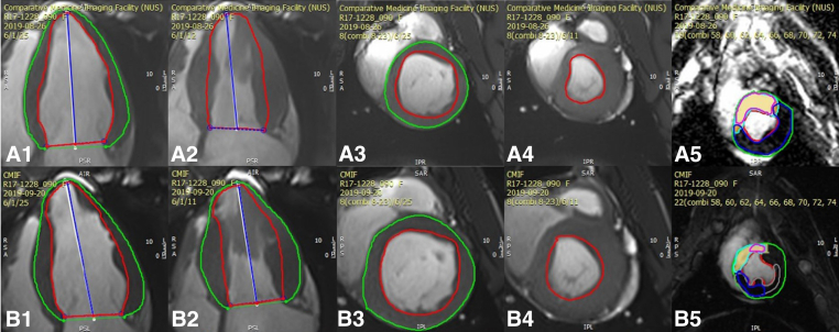
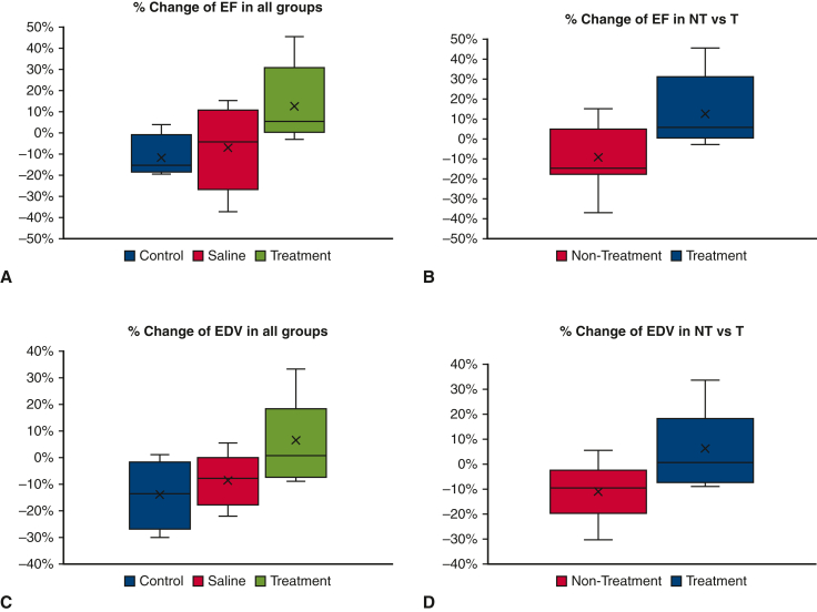
Results: Animals undergoing NOGA-guided hydrogel injections at 8 weeks post-MI demonstrated a significant improvement of the selected left ventricular parameters at a 12-week follow-up. Compared to nonintervention, the hydrogel-based therapy provided significant improvements in end-diastolic volume (-11.0% ± 11.1% vs 6.3% ± 15.2%; P = .008) and ejection fraction (-9.1% ± 16% vs 12.7% ± 18.6%; P = .009). In the slice closest to the apex, significant differences in scar area were observed; the treatment group demonstrated a smaller mean scar area in the infarcted zone compared with the control group (47.1% ± 7.0% vs 59% ± 8.2%; P = .013) and a smaller mean scar area in the border zone compared with the saline group (31.4% ± 8.3% vs 42.6% ± 9.0%; P = .016).
Conclusions: The study implies a translational potential of the hydrogel-based therapy and should trigger clinical trials focused on establishing a restoration therapy that can be integrated into a clinical protocol.
Keywords: NOGA; hydrogel; myocardial infarction; myocardial restoration; transcatheter.
© 2023 The Author(s).
Figures


Similar articles
-
An autologous platelet-rich plasma hydrogel compound restores left ventricular structure, function and ameliorates adverse remodeling in a minimally invasive large animal myocardial restoration model: a translational approach: Vu and Pal "Myocardial Repair: PRP, Hydrogel and Supplements".Biomaterials. 2015 Mar;45:27-35. doi: 10.1016/j.biomaterials.2014.12.013. Epub 2015 Jan 13. Biomaterials. 2015. PMID: 25662492
-
Transcatheter based electromechanical mapping guided intramyocardial transplantation and in vivo tracking of human stem cell based three dimensional microtissues in the porcine heart.Biomaterials. 2013 Mar;34(10):2428-41. doi: 10.1016/j.biomaterials.2012.12.021. Epub 2013 Jan 16. Biomaterials. 2013. PMID: 23332174
-
Citrate-Saline-Formulated mRNA Delivery into the Heart Muscle with an Electromechanical Mapping and Injection Catheter Does Not Lead to Therapeutic Effects in a Porcine Chronic Myocardial Ischemia Model.Hum Gene Ther. 2021 Oct;32(19-20):1295-1307. doi: 10.1089/hum.2021.149. Hum Gene Ther. 2021. PMID: 34494459
-
Cortical Bone Stem Cell Therapy Preserves Cardiac Structure and Function After Myocardial Infarction.Circ Res. 2017 Nov 10;121(11):1263-1278. doi: 10.1161/CIRCRESAHA.117.311174. Epub 2017 Sep 14. Circ Res. 2017. PMID: 28912121 Free PMC article.
-
Clinical aspects of left ventricular diastolic function assessed by Doppler echocardiography following acute myocardial infarction.Dan Med Bull. 2001 Nov;48(4):199-210. Dan Med Bull. 2001. PMID: 11767125 Review.
Cited by
-
Interventional therapies for chronic heart failure: An overview of recent developments.ESC Heart Fail. 2025 Apr;12(2):1081-1094. doi: 10.1002/ehf2.15114. Epub 2024 Nov 11. ESC Heart Fail. 2025. PMID: 39523803 Free PMC article. Review.
References
LinkOut - more resources
Full Text Sources
Miscellaneous
