

Selenium status in term neonates, according to birth weight and gestational age, in relation to maternal hypertensive pathology
- PMID: 37063670
- PMCID: PMC10101720
- DOI: 10.3389/fped.2023.1157689
Selenium status in term neonates, according to birth weight and gestational age, in relation to maternal hypertensive pathology
Abstract
Background: Pregnancy represents a state of increased oxidative stress and antioxidants, in which selenium (Se) plays a pivotal role, contribute to maintain the oxidative balance. If antioxidant defenses are depleted, placental function is disrupted, resulting in pregnancy complications, including pregnancy-induced hypertension (PIH). Little is known about fetal selenium status in concomitant relation to maternal PIH, gestational age (GA) and birthweight (BW).
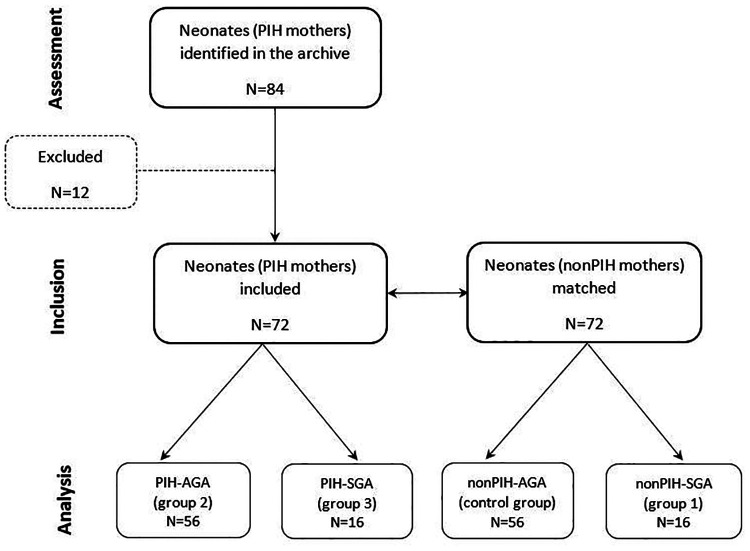
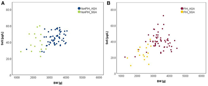
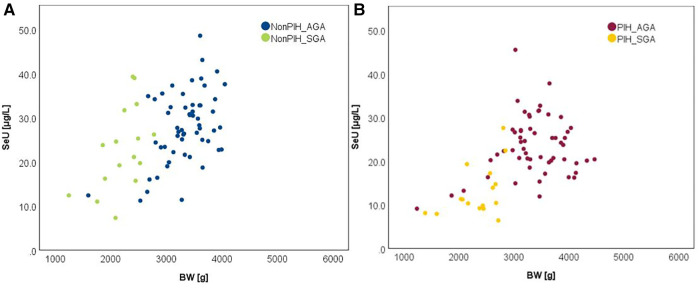
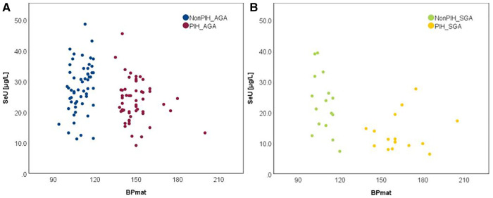
Methods: We examined over a 3-year period the serum (SeS) and urine selenium (SeU) status in term neonates from normotensive (nonPIH) and hypertensive (PIH) mothers as clinical markers of oxidative stress. In this retrospective observational study, 72 neonates with maternal PIH were matched for GA and BW to 72 neonates of normotensive mothers. Four groups were obtained, based on maternal PIH and BW relative to GA (appropriate-for-gestational-age-AGA, small-for-gestational-age-SGA): nonPIH-AGA (control group), nonPIH-SGA, PIH-AGA, and PIH-SGA.
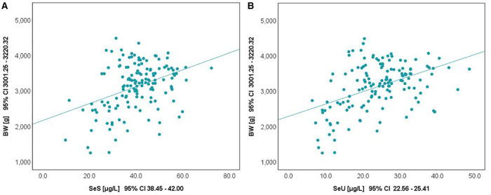
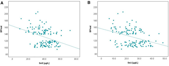
Results: The results showed significant differences (p < 0.001) in selenium levels among the study groups: SeS - 44.85 ± 7.56 μg/L in nonPIH-AGA, 39.62 ± 11.42 μg/L in nonPIH-SGA, 40.01 ± 10.07 μg/L in PIH-AGA, and 25.39 ± 8.99 μg/L in PIH-SGA; SeU - 27.98 ± 7.99 μg/L in nonPIH-AGA, 22.85 ± 9.48 μg/L in nonPIH-SGA, 23.44 ± 6.73 μg/L in PIH-AGA, and 13.05 ± 5.86 μg/L in PIH-SGA. Selenium depletion was more common in neonates born from hypertensive mothers and those born small for gestational age. Though moderate in intensity, selenium levels were positively correlated with BW (0.319 for SeS, 0.397 for SeU) and negatively correlated with maternal systolic blood pressure (-0.313 for SeS, -0.324 for SeU). The main independent effects on SeS and SeU of each maternal blood pressure and birth weight turned out statistically significant. In interaction, a more pronounced effect was reached in PIH-SGA neonates.
Conclusion: Selenium status seemed to reflect the negative impact that PIH exerts in neonates during intrauterine development. Clinical markers of selenium status could thus be of great value for tracking responses of individuals to selenium supplementation as part of health improvement and harm mitigation approaches.
Keywords: birthweight; gestational age; neonate; oxidative stress; pregnancy-induced hypertension; selenium.
© 2023 Bizerea-Moga, Pitulice, Bizerea-Spiridon, Angelescu, Marginean and Moga.
Conflict of interest statement
The authors declare that the research was conducted in the absence of any commercial or financial relationships that could be construed as a potential conflict of interest.
Figures

Similar articles
-
Pregnancy Induced Hypertension Versus Small Weight for Gestational Age: Cause of Neonatal Hematological Disorders.Clin Lab. 2018 Jul 1;64(7):1241-1248. doi: 10.7754/Clin.Lab.2018.180302. Clin Lab. 2018. PMID: 30146849
-
Placental morphological features of small for gestational age preterm neonates born to mothers with pregnancy-induced hypertension.Front Pediatr. 2023 Mar 21;11:1093622. doi: 10.3389/fped.2023.1093622. eCollection 2023. Front Pediatr. 2023. PMID: 37025291 Free PMC article.
-
Mid-pregnancy maternal leptin levels, birthweight for gestational age and preterm delivery.Clin Endocrinol (Oxf). 2013 Apr;78(4):607-13. doi: 10.1111/cen.12029. Clin Endocrinol (Oxf). 2013. PMID: 22934578 Free PMC article.
-
Retinopathy of Prematurity: Incidence and Risk Factor Analysis in Small for Gestational Age Neonates Compared to Appropriate for Gestational Age.J Pharm Bioallied Sci. 2023 Jul;15(Suppl 2):S1266-S1269. doi: 10.4103/jpbs.jpbs_130_23. Epub 2023 Jul 11. J Pharm Bioallied Sci. 2023. PMID: 37694017 Free PMC article.
-
Pregnancy-Induced Hypertension Pathophysiology and Contemporary Management Strategies: A Narrative Review.Cureus. 2024 Jul 6;16(7):e63961. doi: 10.7759/cureus.63961. eCollection 2024 Jul. Cureus. 2024. PMID: 39105037 Free PMC article. Review.
Cited by
-
Is Maternal Selenium Status Associated with Pregnancy Outcomes in Physiological and Complicated Pregnancy?Nutrients. 2024 Aug 27;16(17):2873. doi: 10.3390/nu16172873. Nutrients. 2024. PMID: 39275191 Free PMC article.
References
LinkOut - more resources
Full Text Sources
Miscellaneous