. 2023 Mar 31:14:1098140.
doi: 10.3389/fimmu.2023.1098140. eCollection 2023.

Identifying TNF and IL6 as potential hub genes and targeted drugs associated with scleritis: A bio-informative report

Affiliations

Identifying TNF and IL6 as potential hub genes and targeted drugs associated with scleritis: A bio-informative report

Feiyue Yan et al. Front Immunol. .

Abstract

Background: Scleritis is a serious inflammatory eye disease that can lead to blindness. The etiology and pathogenesis of scleritis remain unclear, and increasing evidence indicates that some specific genes and proteins are involved. This study aimed to identify pivotal genes and drug targets for scleritis, thus providing new directions for the treatment of this disease.

Methods: We screened candidate genes and proteins associated with scleritis by text-mining the PubMed database using Python, and assessed their functions by using the DAVID database. Gene Ontology (GO) and Kyoto Encyclopedia of Genes and Genomes (KEGG) analyses were used to identify the functional enrichment of these genes and proteins. Then, the hub genes were identified with CytoHubba and assessed by protein-protein interaction (PPI) network analysis. And the serum from patients with active scleritis and healthy subjects were used for the validation of hub genes. Finally, the DGIdb database was used to predict targeted drugs for the hub genes for treating scleritis.

Results: A total of 56 genes and proteins were found to be linked to scleritis, and 65 significantly altered pathways were identified in the KEGG analysis (FDR < 0.05). Most of the top five pathways involved the categories "Rheumatoid arthritis," "Inflammatory bowel disease", "Type I diabetes mellitus," and "Graft-versus-host disease". TNF and IL6 were considered to be the top 2 hub genes through CytoHubba. Based on our serum samples, hub genes are expressed at high levels in active scleritis. Five scleritis-targeting drugs were found among 88 identified drugs.

Conclusions: This study provides key genes and drug targets related to scleritis through bioinformatics analysis. TNF and IL6 are considered key mediators and possible drug targets of scleritis. Five drug candidates may play an important role in the diagnosis and treatment of scleritis in the future, which is worthy of the further experimental and clinical study.

Keywords: IL6; PPI network; TNF; drug targets; hub genes; scleritis.


Conflict of interest statement

The authors declare that the research was conducted in the absence of any commercial or financial relationships that could be construed as a potential conflict of interest.

Figures

Figure 1
Figure 1
Overview of research findings. (A)Number of articles published from 1910 to February 2022. (B) Percentage of article categories. (C) Word cloud of the 100 most frequently used words in the literature abstract (the larger the font size, the higher the frequency).
Figure 2
Figure 2
Flow chart for the selection of candidate genes and proteins.
Figure 3
Figure 3
GO analysis bubble map of scleritis-associated genes. (A) Cellular components (CC); (B) Molecular functions (MF); (C) Biological process (BP). The circles highlighted in purple had higher FDRs, while those highlighted in yellow had lower FDRs. X-axis stands for the Gene Ratio of the pathway. Counts of pathways are represented by circles, and a larger circle indicates a pathway with a higher enrichment. (Crafting website: https://www.chiplot.online/).
Figure 4
Figure 4
Bubble map of scleritis-associated genes for the top ten pathways of KEGG. X-axis stands for the Gene Ratio of the pathway. (Crafting website: https://www.chiplot.online/).
Figure 5
Figure 5
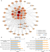
PPI network and KEGG pathway analysis for key clusters. (A) Construction and analysis of the PPI network. Hexagons represent cluster 1, diamonds represent cluster 2, and other circles showed interactions between multiple genes in the PPI network. The size and color of the graph are related to the degree. The bigger and more red, the higher degree on the map. (B) KEGG pathway analysis for cluster 1; (C) KEGG pathway analysis for cluster 2.
Figure 6
Figure 6
Screening for hub genes. (A) Hub genes are obtained in Venn analysis. (B) The scleritis-associated genes PPI network was constructed by Cytohubba with 7 hub genes and 45 other genes. Nodes in pink diamonds represent the top 7 hub genes, and other blue circles represent the other 45 genes, indicating gene interactions. (C) Coexpression analysis of seven hub genes in Homo sapiens.
Figure 7
Figure 7
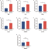
Validation of the relative protein expression levels in serum. (A-G) The levels of TNF, IL6, IFNγ, IL1β, ICAM1, IL17A and MMP9 in patients with active scleritis (n = 5) and healthy control subjects (n = 5). *P < 0.05; **P < 0.01; NS, no significant (two-tailed student’s t-test). Graphs show mean ± SEM.
Figure 8
Figure 8
Interaction network between hub genes and targeted drugs. TNF and IL6, the two drug-targeted genes, are represented as pink diamonds. As identified therapeutic agents, 88 drugs were displayed in triangles and regularly arranged according to their score level; triangles closer to the center have higher scores. DALIMUMAB, ETANERCEPT, INFLIXIMAB, INSULIN, PF-04236921 represented by a hexagon. Drugs approved by FDA are shown in purple, while those not approved are shown in gray.

References

    1. Okhravi N, Odufuwa B, McCluskey P, Lightman S. Scleritis. Survey Ophthalmol (2005) 50:351–63. doi: 10.1016/j.survophthal.2005.04.001 - DOI - PubMed
    1. Watson P, Romano A. The impact of new methods of investigation and treatment on the understanding of the pathology of scleral inflammation. Eye (London England) (2014) 28:915–30. doi: 10.1038/eye.2014.110 - DOI - PMC - PubMed
    1. Samalia P, Sims J, Niederer R. Necrotizing syphilitic scleritis: The resurgence of syphilis in new Zealand. Asia-Pac J Ophthalmol (Philadelphia Pa.) (2021) 10:417. doi: 10.1097/apo.0000000000000383 - DOI - PubMed
    1. Tsui E, Sarrafpour S, Modi YS. Posterior scleritis with choroidal effusion secondary to herpes zoster ophthalmicus. Ocular Immunol Inflammation (2018) 26:184–6. doi: 10.1080/09273948.2017.1351572 - DOI - PubMed
    1. Artifoni M, Rothschild PR, Brézin A, Guillevin L, Puéchal X. Ocular inflammatory diseases associated with rheumatoid arthritis. Nat Rev Rheumatol (2014) 10:108–16. doi: 10.1038/nrrheum.2013.185 - DOI - PubMed

Publication types