. 2023 Mar 31:14:1139358.
doi: 10.3389/fimmu.2023.1139358. eCollection 2023.

Uncovering a novel role of focal adhesion and interferon-gamma in cellular rejection of kidney allografts at single cell resolution

Affiliations

Uncovering a novel role of focal adhesion and interferon-gamma in cellular rejection of kidney allografts at single cell resolution

Ahmad Halawi et al. Front Immunol. .

Abstract

Background: Kidney transplant recipients are currently treated with nonspecific immunosuppressants that cause severe systemic side effects. Current immunosuppressants were developed based on their effect on T-cell activation rather than the underlying mechanisms driving alloimmune responses. Thus, understanding the role of the intragraft microenvironment will help us identify more directed therapies with lower side effects.

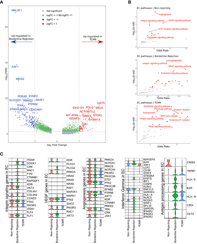
Methods: To understand the role of the alloimmune response and the intragraft microenvironment in cellular rejection progression, we conducted a Single nucleus RNA sequencing (snRNA-seq) on one human non-rejecting kidney allograft sample, one borderline sample, and T-cell mediated rejection (TCMR) sample (Banff IIa). We studied the differential gene expression and enriched pathways in different conditions, in addition to ligand-receptor (L-R) interactions.

Results: Pathway analysis of T-cells in borderline sample showed enrichment for allograft rejection pathway, suggesting that the borderline sample reflects an early rejection. Hence, this allows for studying the early stages of cellular rejection. Moreover, we showed that focal adhesion (FA), IFNg pathways, and endomucin (EMCN) were significantly upregulated in endothelial cell clusters (ECs) of borderline compared to ECs TCMR. Furthermore, we found that pericytes in TCMR seem to favor endothelial permeability compared to borderline. Similarly, T-cells interaction with ECs in borderline differs from TCMR by involving DAMPS-TLRs interactions.

Conclusion: Our data revealed novel roles of T-cells, ECs, and pericytes in cellular rejection progression, providing new clues on the pathophysiology of allograft rejection.

Keywords: alloimmunity; cellular microenvironment; graft rejection; kidney transplantation; single-cell analysis.


Conflict of interest statement

KV is employed by Q32 Bio Inc. The remaining authors declare that the research was conducted in the absence of any commercial or financial relationships that could be construed as a potential conflict of interest.

Figures

Figure 1
Figure 1
(A–C) Umaps of non-rejecting, borderline, and T-cell mediated rejection (TCMR) samples showing different clusters. (D) Umaps showing the expression of certain canonical markers used for cluster annotation. PT (Proximal tubules), AL-1 (Ascending loop of Henle-1), AL-2 (Ascending loop of Henle-2), CNT-PC (Connecting tubules-Principal cells), DCT-PC-CNT (Distal convoluted tubules-Principal cells- Connecting tubules), DL (Descending loop of Henle), IC-A (intercalated cells A), IC-B (intercalated cells-B), DCT (Distal convoluted tubules), EC (Endothelial cells), PO (podocytes).
Figure 2
Figure 2
(A) Plot showing the differentially expressed genes between borderline (red) and TCMR (blue). (B) Pathway analysis of T-cell cluster in borderline compared to TCMR samples using Hallmark database showed enrichment for interferon-gamma and alpha responses and allograft rejection pathways (C) Violin plots showing the level expression of genes part of allograft rejection and interferon-gamma response pathways in three conditions.
Figure 3
Figure 3
(A) Volcano plot showing the up-regulated genes in endothelial cell clusters (ECs) between borderline and TCMR. Blue and red dots and genes up-regulated in borderline and TCMR, respectively; green dots are genes with log-fold change (logFC) between 1 and -1; gray dots are genes that did not reach a significance level. (B) Scatter plots showing the top enriched pathways in non-rejection, borderline, and TCMR samples. (C) Violin plots showing the level expression of genes part of integrin signaling, VEGF, angiogenesis, interferon-gamma response, and antigen processing pathways in ECs clusters in the three conditions.
Figure 4
Figure 4
(A) T-cells, endothelial cells (EC), and pericytes interactions. Focal adhesion represents the interaction between ECs and the surrounding basement membrane (BM); adhesion plaque is the interaction between ECs and pericytes. (B) Violin plot showing canonical markers expressed by pericyte clusters in borderline and TCMR. (C, D) Circos plot showing the ligand-receptor interactions between pericytes in yellow and endothelial cells in orange in borderline and TCMR; the heads of arrows are toward the receptor side; the halo color around the arrow represents the strength of this interaction as per L-R score. (E, F) Circos plot showing the ligand-receptor interactions between T-cells in yellow and endothelial cells in orange in borderline and TCMR; the heads of arrows are toward the receptor side; the halo color around the arrow represents the strength of this interaction as per L-R score.

References

    1. Cooper JE. Evaluation and treatment of acute rejection in kidney allografts. Clin J Am Soc Nephrol (2020) 15(3):430–8. doi: 10.2215/CJN.11991019 - DOI - PMC - PubMed
    1. Wu H, Malone AF, Donnelly EL, Kirita Y, Uchimura K, Ramakrishnan SM, et al. . Single-cell transcriptomics of a human kidney allograft biopsy specimen defines a diverse inflammatory response. J Am Soc Nephrol (2018) 29(8):2069–80. doi: 10.1681/ASN.2018020125 - DOI - PMC - PubMed
    1. Liao J, Yu Z, Chen Y, Bao M, Zou C, Zhang H, et al. . Single-cell RNA sequencing of human kidney. Sci Data (2020) 7(1):4. doi: 10.1038/s41597-019-0351-8 - DOI - PMC - PubMed
    1. Ding J, Adiconis X, Simmons SK, Kowalczyk MS, Hession CC, Marjanovic ND, et al. . Systematic comparison of single-cell and single-nucleus RNA-sequencing methods. Nat Biotechnol (2020) 38(6):737–46. doi: 10.1038/s41587-020-0465-8 - DOI - PMC - PubMed
    1. Wu H, Kirita Y, Donnelly EL, Humphreys BD. Advantages of single-nucleus over single-cell RNA sequencing of adult kidney: Rare cell types and novel cell states revealed in fibrosis. J Am Soc Nephrol (2019) 30(1):23–32. doi: 10.1681/ASN.2018090912 - DOI - PMC - PubMed

Publication types