. 2023 Mar 31:14:1130933.
doi: 10.3389/fimmu.2023.1130933. eCollection 2023.

ILC2 require cell-intrinsic ST2 signals to promote type 2 immune responses

Affiliations

ILC2 require cell-intrinsic ST2 signals to promote type 2 immune responses

Patrycja M Topczewska et al. Front Immunol. .

Abstract

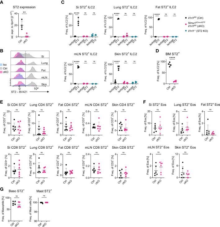
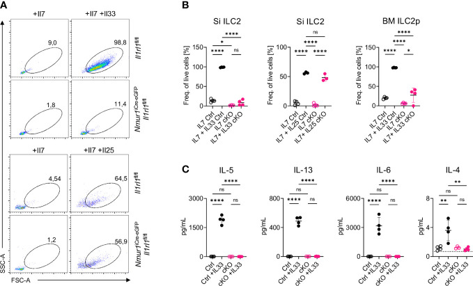
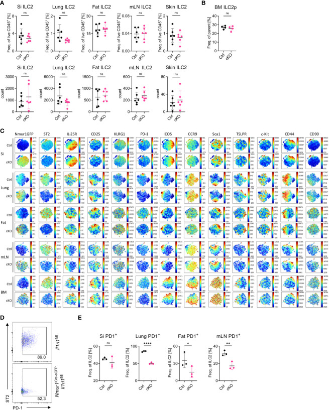
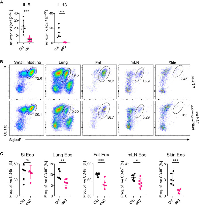
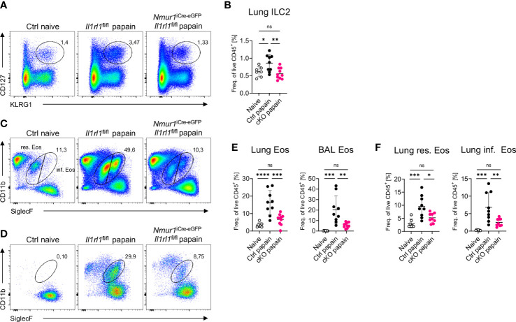
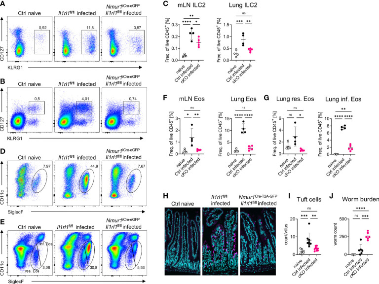
The initiation of type 2 immune responses at mucosal barriers is regulated by rapidly secreted cytokines called alarmins. The alarmins IL-33, IL-25 and TSLP are mainly secreted by stromal and epithelial cells in tissues and were linked to chronic inflammatory diseases, such as allergic lung inflammation, or to resistance against worm infections. Receptors for alarmins are expressed by a variety of immune cells, including group 2 innate lymphoid cells (ILC2s), an early source of the type 2 cytokines, such as IL-5 and IL-13, which have been linked to atopic diseases and anti-worm immunity as well. However, the precise contribution of the IL-33 receptor signals for ILC2 activation still needs to be completed due to limitations in targeting genes in ILC2. Using the newly established Nmur1 iCre-eGFP mouse model, we obtained specific conditional genetic ablation of the IL-33 receptor subunit ST2 in ILC2s. ST2-deficient ILC2s were unresponsive to IL-33 but not to stimulation with the alarmin IL-25. As a result of defective ST2 signals, ILC2s produced limited amounts of IL-5 and IL-13 and failed to support eosinophil homeostasis. Further, ST2-deficient ILC2s were unable to expand and promote the recruitment of eosinophils during allergic lung inflammation provoked by papain administration. During infection with Nippostrongylus brasiliensis, ILC2-intrinsic ST2 signals were required to mount an effective type 2 immune response against the parasite leading to higher susceptibility against worm infection in conditional knockout mice. Therefore, this study argues for a non-redundant role of cell-intrinsic ST2 signals triggering proper activation of ILC2 for initiation of type 2 immunity.

Keywords: IL-33 and ST2; ILC2 - group 2 innate lymphoid cell; innate immunity; mucosal immunity; type 2 immune response.


Conflict of interest statement

DA has contributed to scientific advisory boards at Pfizer, Takeda, FARE and the KRF. The remaining authors declare that the research was conducted in the absence of any commercial or financial relationships that could be construed as a potential conflict of interest. The handling editor HK declared a past co-authorship with the author DA.

Figures

Figure 1
Figure 1
A model for ILC2-specific genetic ablation of ST2. (A), Expression of the Il1rl1 gene in Il1rl1 flox/flox (Ctrl) and Nmur1 iCre-eGFP Il1rl1 flox/flox (cKO) sort-purified ILC2s. (B), Flow cytometric histograms of ST2 expression in ILC2s of Il1rl1 flox/flox (Ctrl) and Nmur1 iCre-eGFP Il1rl1 flox/flox (cKO) mice across organs, including isotype control on Il1rl1 flox/flox (Iso) in the steady state. ILC2s were gated on live CD45+ Lin- (CD3, CD5, CD19, Ly6G, Fcϵr1), CD127+ and KLRG1+. (C–G) Percentage of ST2+ expression in Il1rl1 flox/flox (Ctrl), Nmur1 iCre-eGFP Il1rl1 flox/flox (cKO) (D–G) and Il1rl1 -/- (ST2 KO) mice including isotype control (C) across different organs in the indicated immune cells. Basophils from spleen and mast cells from the peritoneal lavage. For (C–G): Each symbol represents data from one mouse, data are representative of two experiments with four to six mice per group. Mean +/- SD, Student’s t-Test. ns, not significant, **p < 0.01, ****p < 0.0001.
Figure 2
Figure 2
ILC2 of Nmur1 iCre-eGFP Il1rl1 flox/flox mice are unresponsive to IL-33. (A), Flow cytometric plots of small intestine ILC2s from Il1rl1 flox/flox and Nmur1 iCre-eGFP Il1rl1 flox/flox mice after stimulation with IL-7, IL-7 and IL-33 or IL-7 and IL-25. ILC2s were gated on live CD45+ Lin- (CD3, CD5, CD19, Ly6G, Fcer1), CD127+ and KLRG1+. (B), Quantification of ILC2 activation from (A) and bone marrow ILC2s of Il1rl1 flox/flox (Ctrl) and Nmur1 iCre-eGFP Il1rl1 flox/flox (cKO) mice after a three day culture with cytokines IL-7 or IL-7, IL-33 or IL-7, IL-25. (C), Concentration of the indicated cytokine as determined in the cell culture supernatant 3 days after stimulation as in (A, B). For (B, C): Each symbol represents data from one mouse, data are representative of two experiments with four mice per group. Mean +/- SD, One-way ANOVA with multiple comparison. ns, not significant, *p < 0.05, **p < 0.01, ****p < 0.0001.
Figure 3
Figure 3
ILC2 develop in normal proportions without ST2 but have reduced PD-1 expression (A), Quantification of relative and absolute ILC2 numbers in Il1rl1 flox/flox (Ctrl) and Nmur1 iCre-eGFP Il1rl1 flox/flox (cKO) mice across different organs in steady state. (B), Quantification of relative ILC2 progenitor numbers in Il1rl1 flox/flox (Ctrl) and Nmur1 iCre-eGFP Il1rl1 flox/flox (cKO) mice in the BM. (C), t-distributed stochastic neighbour embedding plots showing the expression level of different ILC2 markers. ILC2s from naïve Il1rl1 flox/flox (Ctrl) and Nmur1 iCre-eGFP Il1rl1 flox/flox (cKO) mice are shown. Each dot represents a single cell. Data are representative of two experiments with four to five mice per group. (D), Flow cytometric plots of PD-1 expression in lung ILC2s from Nmur1 iCre-eGFP Il1rl1 flox/flox and Il1rl1 flox/flox. (E), Quantification of PD-1 expression in Il1rl1 flox/flox (Ctrl) and Nmur1 iCre-eGFP Il1rl1 flox/flox (cKO) mice across different organs in steady state. For (A, B, D): Each symbol represents data from one mouse, data are representative of two experiments with three to eight mice per group. Mean +/- SD, student’s t-Test. ns not significant, *p < 0.05, **p < 0.01, ****p < 0.0001.
Figure 4
Figure 4
Loss of constant ST2 signaling results in reduced eosinophil counts. (A), Relative expression of the cytokine IL-5 and IL-13 of Il1rl1 flox/flox (Ctrl) and Nmur1 iCre-eGFP Il1rl1flox/flox (cKO) mice from the lung at steady state. (B), Flow cytometric plots of eosinophils from Il1rl1 flox/flox and Nmur1 iCre-eGFP Il1rl1 flox/flox mice across different organs in steady state. Eosinophils were gated on live CD45+ Lin- (CD3, CD5, CD19), CD11b+ and SiglecF+. (C), Quantification of (B) in Il1rl1 flox/flox (Ctrl) and Nmur1 iCre-eGFP Il1rl1 flox/flox (cKO) mice across different organs in steady state. For (A, C): Each symbol represents data from one mouse, data are representative of two experiments with five to seven mice per group. Mean +/- SD, student’s t-Test. ns, not significant, *p < 0.05, **p < 0.01, ***p < 0.001.
Figure 5
Figure 5
ST2-deficient ILC2s fail to recruit eosinophils in acute lung inflammation. (A, C, D), Flow-cytometric plots showing ILC2s (A) in the Lung, eosinophils (C) in the lung or BAL (D) of untreated Ctrl (naïve) animals and papain treated Nmur1 iCre-eGFP Il1rl1 flox/flox mice (cKO) and littermate controls (Ctrl). Data are representative of two experiments with four to six mice per group. (B, E), Quantification of relative ILC2 (B) and eosinophil numbers from the lung or BAL (E) in the lung and BAL. (F), Quantification of relative resident and inflammatory eosinophil numbers in the lung. For (B, E, F): One-way ANOVA with multiple comparisons, ns, not significant, *p < 0.05, **p < 0.01, ***p < 0.001, ****p < 0.0001.
Figure 6
Figure 6
ILC2 intrinsic ST2 is required for immunity against N. brasiliensis infection. (A, B), Flow-cytometric plots showing ILC2s in the mLN (A) and in the lung (B) of untreated Ctrl (naïve) animals and infected Nmur1 iCre-eGFP Il1rl1 flox/flox mice (cKO) and littermate controls (Ctrl). Mice were infected with N. brasiliensis. (C), Quantification of (A, B). (D, E) Flow-cytometric plots showing eosinophils in the mLN (D) and in the lung (E) of untreated Ctrl (naïve) animals and infected Nmur1 iCre-eGFP Il1rl1 flox/flox mice (cKO) and littermate controls (Ctrl). Mice were infected with N. brasiliensis. (F), Quantification of (D) and (E, G), Quantification of relative resident and inflammatory eosinophil numbers in the lung of untreated Ctrl (naïve) animals and infected Nmur1 iCre-eGFP Il1rl1 flox/flox mice (cKO) and littermate controls (Ctrl). (H), Immunofluorescence migrographs of histological sections of the murine small intestine, tuft cells stained with DCLK1 (magenta), DAPI (blue). (I), Quantification of (H). Tuft cell numbers per villus. Data combined from two independent experiments. (J), Worm burden in untreated animals (naïve) and infected Nmur1 iCre-eGFP Il1rl1 flox/flox mice (cKO) or littermate controls (Ctrl). For (C, F, G, I, J): Data are representative of two experiments with four to six mice per group. One-way ANOVA with multiple comparisons, ns, not significant, *p < 0.05, **p < 0.01, ***p < 0.001, ****p < 0.0001.

References

    1. Stanbery AG, Smita S, von Moltke J, Tait Wojno ED, Ziegler SF. TSLP, IL-33, and IL-25: Not just for allergy and helminth infection. J Allergy Clin Immunol (2022) 150:1302–13. doi: 10.1016/j.jaci.2022.07.003 - DOI - PMC - PubMed
    1. Mahlakoiv T, Flamar AL, Johnston LK, Moriyama S, Putzel GG, Bryce PJ, et al. . Stromal cells maintain immune cell homeostasis in adipose tissue via production of interleukin-33. Sci Immunol (2019) 4(35). doi: 10.1126/sciimmunol.aax0416 - DOI - PMC - PubMed
    1. Bonilla WV, Frohlich A, Senn K, Kallert S, Fernandez M, Johnson S, et al. . The alarmin interleukin-33 drives protective antiviral CD8(+) T cell responses. Science (2012) 335:984–9. doi: 10.1126/science.1215418 - DOI - PubMed
    1. Rana BMJ, Jou E, Barlow JL, Rodriguez-Rodriguez N, Walker JA, Knox C, et al. . A stromal cell niche sustains ILC2-mediated type-2 conditioning in adipose tissue. J Exp Med (2019) 216:1999–2009. doi: 10.1084/jem.20190689 - DOI - PMC - PubMed
    1. Spallanzani RG, Zemmour D, Xiao T, Jayewickreme T, Li C, Bryce PJ, et al. . Distinct immunocyte-promoting and adipocyte-generating stromal components coordinate adipose tissue immune and metabolic tenors. Sci Immunol (2019) 4(35). doi: 10.1126/sciimmunol.aaw3658 - DOI - PMC - PubMed

Publication types