Case Reports
. 2023 Apr 12;2023(4):rjad172.
doi: 10.1093/jscr/rjad172. eCollection 2023 Apr.

Giant retroperitoneal leiomyosarcoma: a case report

Affiliations
Case Reports

Giant retroperitoneal leiomyosarcoma: a case report

Amine Hermi et al. J Surg Case Rep. .

Abstract

Retroperitoneal leiomyosarcomas are rare tumors, mostly malignant. They are silent slow growing, and at the time of diagnosis, they are often of a considerable size. Management necessitates en bloc resection of the mass with adjacent organs, which is often challenging due to large size of the tumor. Herein, we present a case of 59-year-old male patient presenting for surgical management of 190 × 150 × 140 mm retroperitoneal leiomyosarcoma.

Keywords: leiomyosarcoma; retroperitoneal; sarcoma.


Conflict of interest statement

The authors declare that they have no competing interests.

Figures

Figure 1
Figure 1
Axial (A) and frontal (B) view of Computed tomography scan showing a large right retroperitoneal solid mass, with lobulated thickened wall measuring 190 × 140 × 120 mm, heterogeneously enhancing, displacing anteriorly duodenal loops, laterally the right colon, medially the inferior vena cava and abdominal aorta.
Figure 2
Figure 2
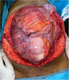
Preoperative view of the large lobulated mass occupying largely the abdominal cavity.
Figure 3
Figure 3
(A) Microscopic examination showing spindle cells with elongated hyperchromatic nuclei and arranged in interlacing bundles of (HE X 200). (B) Tumor cells showing nuclear atypia and eosinophilic cytoplasm (HE X 400).

References

    1. Marko J, Wolfman DJ. Retroperitoneal leiomyosarcoma from the radiologic pathology archives. Radiographics 2018;38:1403–20. - PMC - PubMed
    1. Dull BZ, Smith B, Tefera G. Surgical management of retroperitoneal leiomyosarcoma arising from the inferior vena cava. Weber S J Gastrointest Surg 2013;17:2166–71. - PMC - PubMed
    1. Matsuoka T, Tsujioka H, Matsuoka S, Sorano S, et al. Retroperitoneal leiomyosarcoma: a case report. Eur J Gynaecol Oncol 2017;38:150–2. - PubMed
    1. Olimpiadi Y, Song S, Hu JS, Matcuk GR, et al. Contemporary management of retroperitoneal soft tissue sarcomas. Curr Oncol Rep 2015;17:39. - PubMed
    1. Von Mehren M, Kane JM, Agulnik M, Bui MM, et al. Soft tissue sarcoma, version 2.2022, NCCN clinical practice guidelines in oncology. J Natl Compr Cancer Netw JNCCN 2022;20:815–33. - PMC - PubMed

Publication types