. 2023 Mar 31:17:1139737.
doi: 10.3389/fnbeh.2023.1139737. eCollection 2023.

Lateral septal nucleus, dorsal part, and dentate gyrus are necessary for spatial and object recognition memory, respectively, in mice

Affiliations

Lateral septal nucleus, dorsal part, and dentate gyrus are necessary for spatial and object recognition memory, respectively, in mice

Ying-Ke Jiang et al. Front Behav Neurosci. .

Abstract

Introduction: Cognitive impairment includes the abnormality of learning, memory and judgment, resulting in severe learning and memory impairment and social activity impairment, which greatly affects the life quality of individuals. However, the specific mechanisms underlying cognitive impairment in different behavioral paradigms remain to be elucidated.

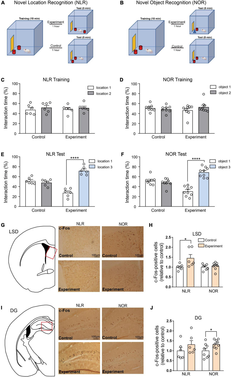
Methods: The study utilized two behavioral paradigms, novel location recognition (NLR) and novel object recognition (NOR), to investigate the brain regions involved in cognitive function. These tests comprised two phases: mice were presented with two identical objects for familiarization during the training phase, and a novel (experiment) or familiar (control) object/location was presented during testing. Immunostaining quantification of c-Fos, an immediate early gene used as a neuronal activity marker, was performed in eight different brain regions after the NLR or NOR test.

Results: The number of c-Fos-positive cells was significantly higher in the dorsal part of the lateral septal nucleus (LSD) in the NLR and dentate gyrus (DG) in the NOR experiment group than in the control group. We further bilaterally lesioned these regions using excitotoxic ibotenic acid and replenished the damaged areas using an antisense oligonucleotide (ASO) strategy.

Discussion: These data reinforced the importance of LSD and DG in regulating spatial and object recognition memory, respectively. Thus, the study provides insight into the roles of these brain regions and suggests potential intervention targets for impaired spatial and object recognition memory.

Keywords: c-Fos; dentate gyrus; dorsal part; lateral septal nucleus (LSD); lesion; recognition memory.


Conflict of interest statement

The authors declare that the research was conducted in the absence of any commercial or financial relationships that could be construed as a potential conflict of interest.

Figures

FIGURE 1
FIGURE 1
Timeline of the study and experimental design. ASO, antisense oligonucleotide; NSO, nonsense oligonucleotide; OFT, open field test; NOR, novel object recognition; NLR, novel location recognition; LSD, the dorsal part of the lateral septal nucleus; DG, dentate gyrus; DAB staining, 3,3′-diaminobenzidine staining. Created with https://biorender.com.
FIGURE 2
FIGURE 2
Lateral septal nucleus, dorsal part (LSD), and dentate gyrus (DG) involved in the regulation of novel location recognition (NLR) and novel object recognition (NOR), respectively. (A,B) The diagram of NLR and NOR tests. During the training phase, the mice explored two identical objects for 10 min. During the test phase, 1 h later, one of the objects was changed into a new object/location. The experimental mice were allowed to explore two distinct objects/locations for 5 min each. The control mice were still exposed to the same objects/locations as in the training phase. (C) The interaction time (%) of NLR during the training phase in control and experiment groups (n = 6 for each group). Unpaired t-test: control: t(10) = 0.7176, p = 0.4894; experiment: t(10) = 0.5592, p = 0.5884. (D) The interaction time (%) of NOR during the training phase in the control (n = 8) and experiment group (n = 9). Unpaired t-test: control: t(14) = 0.6287, p = 0.5397; experiment: t(16) = 1.269, p = 0.2227. (E) The interaction time (%) of NLR during the test phase in the control and experiment groups (n = 6 for each group). Unpaired t-test: control: t(10) = 0.9233, p = 0.3776; experiment: t(10) = 9.113, ****p < 0.0001. (F) The interaction time (%) of NOR during the test phase in control (n = 8) and experiment groups (n = 9). Unpaired t-test: control: t(14) = 0.9937, p = 0.3373; experiment: t(16) = 8.233, ****p < 0.0001. (G) Representative images of c-Fos positive cells in the LSD of control and experiment mice in NLR and NOR tests. Scale bar, 100 μm. (H) Quantification of c-Fos positive cells in the LSD of control and experiment mice in NLR and NOR tests (NLR: n = 6; NOR: n = 8). Unpaired t-test: NLR: t(10) = 2.312, *p = 0.0434; NOR: t(14) = 0.7066, p = 0.4914. All the data conformed to normality. (I) Representative images of c-Fos positive cells in the DG of control and experiment mice in NLR and NOR tests. Scale bar, 100 μm. (J) Quantification of c-Fos positive cells in the DG of control and experiment mice in NLR and NOR tests (NLR: n = 6; NOR: n = 8). Unpaired t-test: NLR: t(10) = 1.177, p = 0.2663; NOR: t(15) = 2.202, *p = 0.0437. All the data conformed to normality.
FIGURE 3
FIGURE 3
Lesions of LSD and DG induced by ibotenic acid contributed to deficits in spatial and object recognition memory. (A) Representative images of NeuN-positive cells in the LSD of saline and ibotenic lesioned mice. Scale bar, 100 μm. (B) The number of NeuN positive cells in the LSD of experiment mice. Ibotenic lesioned mice showed decreased neurons in LSD compared with saline mice (saline: n = 4; Ibotenic: n = 6). Unpaired t-test: t(8) = 5.342, ***p = 0.0007. (C) The interaction time (%) of NLR during the training phase in saline (n = 6) and ibotenic lesioned mice (n = 6). Unpaired t-test: saline: t(10) = 0.3271, p = 0.7504; Ibotenic: t(10) = 0.6613, p = 0.5234. (D) The interaction time (%) of NLR during the test phase. Saline mice showed more preference for the novel location compared with the familiar location (n = 6). Unpaired t-test: t(10) = 12.05, ****p < 0.0001. Ibotenic-lesioned mice showed no significant difference between the two locations. Unpaired t-test: t(10) = 0.396, p = 0.7004. (E) Representative images of NeuN-positive cells in the DG of saline and ibotenic lesioned mice. Scale bar, 100 μm. (F) The optical density of NeuN positive cells in the DG of experiment mice. Ibotenic lesioned mice showed decreased neurons in DG compared with saline mice (saline: n = 5; Ibotenic: n = 5). Unpaired t-test: t(8) = 3.111, *p = 0.0144. (G) The interaction time (%) of NOR during the training phase in saline (n = 6) and ibotenic lesioned mice (n = 6). Unpaired t-test: saline: t(10) = 2.020, p = 0.0710; Ibotenic: t(10) = 0.8544, p = 0.4129. (H) The interaction time (%) of NOR during the test phase. Saline mice showed more preference for the novel object compared with the familiar object (n = 6). Unpaired t-test: t(10) = 2.863, *p = 0.023. Ibotenic-lesioned mice showed no significant difference between the two objects. Unpaired t-test: t(10) = 1.680, p = 0.1239.
FIGURE 4
FIGURE 4
Antisense oligonucleotide injection rescued the spatial recognition memory in the lesioned mice by functional neuronal regeneration. (A) Representative images of NeuN-positive cells in the LSD. Scale bar, 100 μm. (B) The number of NeuN positive cells in the LSD (saline/NSO: n = 4; saline/ASO: n = 6; Ibotenic/NSO: n = 6; Ibotenic/ASO: n = 6). Two-way ANOVA: Ibotenic, F(1,18) = 13.38, p = 0.0018; ASO, F(1,18) = 47.77, p < 0.0001; interaction, F(1,18) = 9.635, p = 0.0061; **p < 0.01, ****p < 0.0001 with Bonferroni’s post-hoc test. (C) The interaction time (%) of NLR during the training phase 15 days after ASO injection (saline/NSO: n = 5; saline/ASO: n = 6; Ibotenic/NSO: n = 6; Ibotenic/ASO: n = 6). Unpaired t-test: saline/NSO: t(8) = 1.272, p = 0.2390; saline/ASO: t(10) = 1.693, p = 0.1213; Ibotenic/NSO: t(10) = 0.9252, p = 0.3766; Ibotenic/ASO: t(10) = 0.5864, p = 0.5706. (D) The interaction time (%) of NLR during the test phase 15 days after ASO injection (saline/NSO: n = 5; saline/ASO: n = 6; Ibotenic/NSO: n = 6; Ibotenic/ASO: n = 6). Unpaired t-test: saline/NSO: t(8) = 6.123, ***p = 0.0003; saline/ASO: t(10) = 4.119, **p = 0.0021; Ibotenic/NSO: t(10) = 0.3960, p = 0.7004; Ibotenic/ASO: t(10) = 4.418, **p = 0.0013. (E) The interaction time (%) of NLR during the training phase 30 days after ASO injection (saline/NSO: n = 5; saline/ASO: n = 6; Ibotenic/NSO: n = 5; Ibotenic/ASO: n = 6). Unpaired t-test: saline/NSO: t(8) = 0.3053, p = 0.7680; saline/ASO: t(10) = 0.005915, p = 0.9954; Ibotenic/NSO: t(8) = 0.2654, p = 0.7974; Ibotenic/ASO: t(10) = 1.225, p = 0.2487. (F) The interaction time (%) of NLR during the test phase 30 days after ASO injection (saline/NSO: n = 5; saline/ASO: n = 6; Ibotenic/NSO: n = 5; Ibotenic/ASO: n = 6). Unpaired t-test: saline/NSO: t(8) = 10.08, ****p < 0.0001; saline/ASO: t(10) = 2.313, *p = 0.0433; Ibotenic/NSO: t(8) = 0.3423, p = 0.7409; Ibotenic/ASO: t(10) = 2.58, *p = 0.0274.
FIGURE 5
FIGURE 5
The adult-born neurons contributed to the improvement of object recognition memory in DG-lesioned mice. (A) Representative images of NeuN-positive cells in the DG. Scale bar, 100 μm. (B) The optical density of NeuN positive cells in the DG (saline/NSO: n = 5; saline/ASO: n = 4; Ibotenic/NSO: n = 4; Ibotenic/ASO: n = 5). Two-way ANOVA: Ibotenic, F(1,14) = 5.367, p = 0.0362; ASO, F(1,14) = 0.5271, p = 0.4798; interaction, F(1,14) = 0.2095, p = 0.6542. (C) The interaction time (%) of NOR during the training phase 15 days after ASO injection (saline/NSO: n = 5; saline/ASO: n = 4; Ibotenic/NSO: n = 4; Ibotenic/ASO: n = 5). Unpaired t-test: saline/NSO: t(8) = 1.305, p = 0.2282; saline/ASO: t(6) = 0.5483, p = 0.6033; Ibotenic/NSO: t(6) = 1.275, p = 0.2495; Ibotenic/ASO: t(8) = 0.4711, p = 0.6502. (D) The interaction time (%) of NOR during the test phase 15 days after ASO injection (saline/NSO: n = 5; saline/ASO: n = 4; Ibotenic/NSO: n = 4; Ibotenic/ASO: n = 5). Unpaired t-test: saline/NSO: t(8) = 4.744, **p = 0.0015; saline/ASO: t(6) = 4.722, **p = 0.0033; Ibotenic/NSO: t(6) = 2.672, *p = 0.0369; Ibotenic/ASO: t(8) = 1.968, p = 0.0847. (E) The interaction time (%) of NOR during the training phase 30 days after ASO injection (saline/NSO: n = 5; saline/ASO: n = 4; Ibotenic/NSO: n = 4; Ibotenic/ASO: n = 5). Unpaired t-test: saline/NSO: t(8) = 0.009471, p = 0.9927; saline/ASO: t(6) = 1.555, p = 0.1710; Ibotenic/NSO: t(6) = 0.7354, p = 0.4898; Ibotenic/ASO: t(8) = 0.6312, p = 0.5455. (F) The interaction time (%) of NOR during the test phase 30 days after ASO injection (saline/NSO: n = 5; saline/ASO: n = 4; Ibotenic/NSO: n = 4; Ibotenic/ASO: n = 5). Unpaired t-test: saline/NSO: t(8) = 2.923, *p = 0.0192; saline/ASO: t(6) = 3.066, *p = 0.0220; Ibotenic/NSO: t(6) = 4.267, **p = 0.0053; Ibotenic/ASO: t(8) = 4.023, **p = 0.0038. (G) Representative images of doublecortin (DCX) positive cells in the DG. Scale bar, 100 μm. (H) The number of DCX-positive cells in the DG (saline/NSO: n = 5; saline/ASO: n = 5; Ibotenic/NSO: n = 5; Ibotenic/ASO: n = 5). Two-way ANOVA: Ibotenic, F(1,16) = 26.67, p < 0.0001; ASO, F(1,16) = 0.5129, p = 0.4842; interaction, F(1,16) = 0.6266, p = 0.4402; *p < 0.05, **p < 0.01 with Bonferroni’s post-hoc test.

References

    1. Aimone J., Li Y., Lee S., Clemenson G., Deng W., Gage F. (2014). Regulation and function of adult neurogenesis: From genes to cognition. Physiol. Rev. 94 991–1026. 10.1152/physrev.00004.2014 - DOI - PMC - PubMed
    1. Ainge J., Heron-Maxwell C., Theofilas P., Wright P., de Hoz L., Wood E. (2006). The role of the hippocampus in object recognition in rats: Examination of the influence of task parameters and lesion size. Behav. Brain Res. 167 183–195. 10.1016/j.bbr.2005.09.005 - DOI - PubMed
    1. An S., Sun H., Wu M., Xie D., Hu S., Ding H., et al. (2021). Medial septum glutamatergic neurons control wakefulness through a septo-hypothalamic circuit. Curr. Biol. 31 1379–1392.e4. 10.1016/j.cub.2021.01.019 - DOI - PubMed
    1. Antunes M., Biala G. (2012). The novel object recognition memory: Neurobiology, test procedure, and its modifications. Cogn. Process. 13 93–110. 10.1007/s10339-011-0430-z - DOI - PMC - PubMed
    1. Berg D. A., Su Y., Jimenez-Cyrus D., Patel A., Huang N., Morizet D., et al. (2019). A common embryonic origin of stem cells drives developmental and adult neurogenesi. Cell 177 654–668.e15. 10.1016/j.cell.2019.02.010 - DOI - PMC - PubMed

LinkOut - more resources