Review
. 2023 Apr 1;13(4):2712-2734.
doi: 10.21037/qims-22-750. Epub 2023 Feb 22.

Advances in differential diagnosis of cerebrovascular diseases in magnetic resonance imaging: a narrative review

Affiliations
Review

Advances in differential diagnosis of cerebrovascular diseases in magnetic resonance imaging: a narrative review

Xin Li et al. Quant Imaging Med Surg. .

Abstract

Background and objective: Cerebrovascular diseases (CVDs), particularly cerebral stroke, remain a primary cause of disability and death worldwide. Accurate diagnosis of CVDs is essential to guide therapeutic decisions and foresee the prognosis. Different CVDs have different pathological processes while they have many signs in common with some other brain diseases. Thus, differential diagnoses of strokes from other primary and secondary CVDs are especially important and challenging.

Methods: This review is composed mainly based on searching PubMed articles between September, 2013 and December 26, 2022 in English.

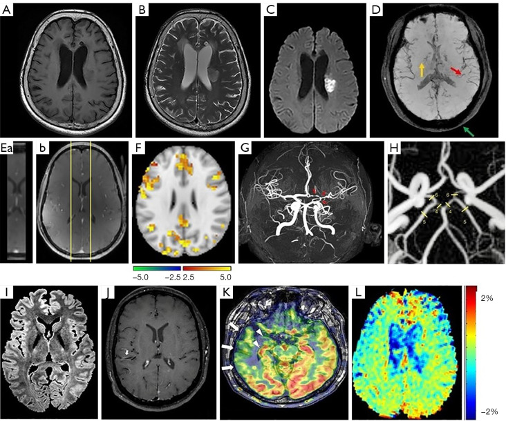
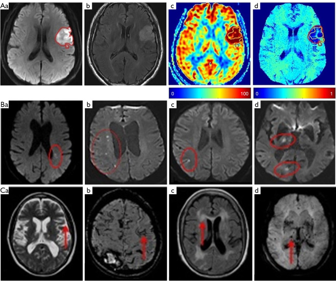
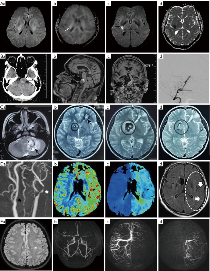
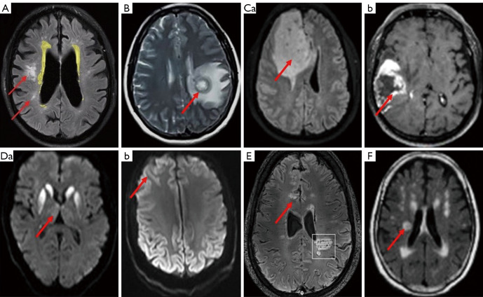
Key content and findings: Neuroimaging is a powerful tool for CVD diagnosis including cerebral angiography, ultrasound, computed tomography, and positron emission tomography as well as magnetic resonance imaging (MRI). MRI excels other imaging techniques by its features of non-invasive, diverse sequences and high spatiotemporal resolution. It can detect hemodynamic, structural alterations of intracranial arteries and metabolic status of their associated brain regions. In acute stroke, differential diagnosis of ischemic from hemorrhagic stroke and other intracranial vasculopathies is a common application of MRI. By providing information about the pathological characteristics of cerebral diseases exhibiting different degrees of behavioral alterations, cognitive impairment, motor dysfunction and other indications, MRI can differentiate strokes from other primary CVDs involving cerebral small vessels and identify vascular dementia from hyponatremia, brain tumors and other secondary or non-primary CVDs.

Conclusions: Recent advances in MRI technology allow clinical neuroimaging to provide unique reference for differentiating many previously inconclusive CVDs. MRI technology is worthy of full exploration while breaking its limitations in clinical applications should be considered.

Keywords: Cerebrovascular diseases (CVDs); cerebral stroke; differential diagnosis; magnetic resonance imaging (MRI); neuroimaging.


Conflict of interest statement

Conflicts of Interest: All authors have completed the ICMJE uniform disclosure form (available at https://qims.amegroups.com/article/view/10.21037/qims-22-750/coif). The authors have no conflicts of interest to declare.

Figures

Figure 1
Figure 1
Basic MRI sequences. (A) T1-weighted-weighted scans. (B) T2-weighted scans. (C) DWI. (A-C). MRI from patient with acute ischemic stroke [reprinted with permission from Lin et al. (23)]. (D) SWI. Some small vessels are marked in red arrows, the red nucleus is marked in yellow arrows, the skull and extracranial lipid are marked in green arrows [reprinted with permission from Qiu et al. (24)]. (E) DCE-MRI. Images of the fast (during contrast) DCE-MRI part at the highest contrast agent concentration (a), and first dynamic phase of the slow (postcontrast) DCE-MRI part after contrast agent arrival (b). The smaller brain area (yellow border lines), planned through the superior sagittal sinus, is acquired for the fast (during contrast) part to obtain shorter dynamic scan intervals [reprinted with permission from de Canjels et al. (21)]. (F) CVR. Brain regions showing significant effects of clinical composite score 1 on the BOLD signal change from the acute to subacute phase of injury [reprinted with permission from Churchill et al. (25)]. The scales are bootstrap ratio. (G) MRA. Time of flight-MRA showing stenotic lesions in the terminal part of internal left carotid, middle and anterior left cerebral arteries (red arrows) in a patient presenting with a secondary central nervous system angiitis in the context of systemic lupus erythematosus [reprinted with permission from Ferlini et al. (26)]. (H) 4D flow MRI. Blood flow is measured in P1 and P2 segments of the posterior cerebral artery [4, 5], and the posterior communicating arteries [6] [reprinted with permission from Malm et al. (27)]. (I) T2/FLAIR. In an axial F section, lower intensity values with a greater degree of myelination [reprinted with permission from Ganzetti et al. (28)]. (J) IVW-MRI. Image shows multiple areas of concentric contrast enhancement in the proximal left and distal right of the middle cerebral arteries (arrows) in a patient presenting with primary angiitis of the central nervous system [reprinted with permission from Ferlini et al. (26)]. (K) ASL-MRI. Interictal ASL shows ipsilateral hypoperfusion in the temporal neocortex (arrows) and in the medial temporal lobe (arrowhead) [reprinted with permission from Sone et al. (29)]. (L) APTw-MRI. Image is from a patient with acute ischemic stroke [reprinted with permission from Lin et al. (23)]. MRI, magnetic resonance imaging; DWI, diffusion-weighted imaging; SWI, susceptibility-weighted imaging; DCE, dynamic contrast-enhanced; CVR, cerebrovascular reactivity; BOLD, blood-oxygen-level dependent; MRA, magnetic resonance angiogram; 4D flow MRI, four-dimensional flow magnetic resonance imaging; FLAIR, fluid-attenuated inversion recovery; IVW, intracranial vessel wall; ASL, arterial spin labeling; APTw, amide-proton-transfer weighted.
Figure 2
Figure 2
MRI illustration of cerebral stroke. (A) Ischemic stroke. Representative images showing the infarct at 58-h in DWI (a, red circle), T2/FLAIR (b), cerebral blood flow (c) and oxygen extraction fraction (d). Reprinted with permission from Wu et al. (53). (B) Lacunar stroke. Stroke topography on DWI in single subcortical (a), multiple subcortical (b), cortical (c), and non-confluent cortical-subcortical regions (d). Reprinted with permission from Tan et al. (54). (C) Hemorrhagic stroke. T2 weighted axial MRI of the brain showing severe cerebral atrophy (a, red arrow); axial MRI of the brain demonstrating a linear gyriform hypointensity (b, red arrow) on SWI sequence in a cortical distribution representative of superficial siderosis; T2/FLAIR weighted MRI showing large confluent lesions (c, red arrow) with a Fazekas 3 rating and maximum intensity projection of SWI sequence of the brain showing multiple diffuse microbleeds (d, red arrow). Reprinted with permission from Karayiannis et al. (55). MRI, magnetic resonance imaging; FLAIR, fluid-attenuated inversion recovery; DWI, diffusion-weighted imaging; SWI, susceptibility-weighted imaging.
Figure 3
Figure 3
MRI illustration of CVDs other than stroke. (A) TIA: images showing no abnormality on the initial DWI scan (a), a hyperintense area in the right basal ganglia on the baseline mean kurtosis map (b, white arrow) and a new ischemic lesion is present in a similar location in the right middle cerebral artery territory on follow-up DWI (c, black arrow) and apparent diffusion coefficient maps (d, white arrowhead) 10 days later, respectively [reprinted with permission from Zhou et al. (76)]. (B) Aneurysm: CT brain axial section showing a dilated basilar artery, suggestive of an aneurysm, compressing the left pons (a, arrow); MRI brain sagittal section showing a partially thrombosed basilar artery aneurysm (b, arrow); MRI brain coronal section showing a dilated basilar artery, and basilar artery aneurysm (c, arrow) and cerebral angiogram confirming a fusosaccular aneurysm arising from V3, V4 segments of left vertebral artery and basilar artery (d, arrow) [reprinted with permission from Bhat et al. (75)]. (C) Malformations. MRI (T1-weighted) detects a lesion located in the left cerebellar hemisphere (a, black arrow) when the patient with cerebral cavernous malformations has the first hemorrhage. At the second hemorrhage, MRI (T2 weighted) shows a lesion in the left frontal lobe (b, which is surgically removed) and a small lesion in right basal ganglia (c and d, black circle) [reprinted with permission from Zhang et al. (77)]. (D) Carotid stenosis. MRA shows stenosis of the proximal internal carotid artery (a, white arrow) and contralateral carotid artery stenosis (a, black arrow). In perfusion-weighted imaging, increased cerebral blood volume (b) and delayed time to peak (c) are shown. FLAIR shows hyperintense vessel signal in FLAIR (d, white arrows) [reprinted with permission from Park et al. (78)]. (E) Moyamoya disease. FLAIR reveals a subacute postischemic lesion in the left-hemispheric white matter (a) and time-of-flight magnetic resonance angiography shows multiple changes of arterial vascular calibers (b). Super-selective 4D ASL-based MRA and digital subtraction angiography of the right-sided ICA (c) and left-sided ICA (d) shows aberrant interhemispheric supply patterns in time-resolved manner [reprinted with permission from Sollmann et al. (79)]. TIA, transient ischemic attack; CVDs, cerebrovascular diseases; DWI, diffusion-weighted imaging; MRI, magnetic resonance imaging; MRA, magnetic resonance angiogram; FLAIR, fluid-attenuated inversion recovery; ASL, arterial spin labeling; ICA, internal carotid artery.
Figure 4
Figure 4
MRI illustration of other brain diseases mimicking CVDs. (A) Hydrocephalus: image showing the periventricular hyperintensities including adjacent changes, but excluding non-adjacent changes in deep white matter. The red arrows mark some of the areas detected as isolated deep white matter changes [reprinted with permission from Snöbohm et al. (115)]. (B) Brain abscess. In the left basal ganglia area, an aberrant, circular space-occupying lesion with long T1 and long T2 signal shadows is present. Its ring wall has relatively even thickness, manifesting as slightly short T1 and T2 signal shadows. The lesion-surrounding area contains patches of apparent edema; the left ventricle displays slight compression-resulted deformation; and the midline structures display minor rightward shift [reprinted with permission from Zhou et al. (118)]. (C) Brain tumors: representative images of patients with astrocytoma (a) and glioblastoma (b) [reprinted with permission from Sartoretti et al. (119)]. (D) Sporadic Creutzfeldt-Jakob disease. DWI shows typical basal ganglia abnormalities (a) and typical features of ‘cortical ribboning’ in the frontal and parietal cortex (b) [reprinted with permission from Rudge et al. (120)]. (E) Multiple sclerosis. 7 T FLAIR-SWI data show several typical white matter hyperintense lesions and global brain atrophy (a) and one large periventricular hyperintense lesion with an encircling hypointense rim (white rectangle, b). Within this lesion, tubular hypointense structures suggestive of veins and circumscribed nodular hypointensities are visible [reprinted with permission from Dal-Bianco et al. (121). (F) Ataxic hemispheres. Image exhibits small, round, high, and central low-signal-intensity lesions in the right frontal lobe and internal capsule in T2/FLAIR [reprinted with permission from Lee et al. (122)]. Red arrows in B-F point to the typical lesions. MRI, magnetic resonance imaging; CVDs, cerebrovascular diseases; DWI, diffusion-weighted imaging; FLAIR, fluid-attenuated inversion recovery; SWI, susceptibility-weighted imaging.

Similar articles

Cited by

References

    1. Salmela MB, Mortazavi S, Jagadeesan BD, Broderick DF, Burns J, et al. ACR Appropriateness Criteria(®) Cerebrovascular Disease. J Am Coll Radiol 2017;14:S34-61. 10.1016/j.jacr.2017.01.051 - DOI - PubMed
    1. Mazzacane F, Mazzoleni V, Scola E, Mancini S, Lombardo I, Busto G, Rognone E, Pichiecchio A, Padovani A, Morotti A, Fainardi E. Vessel Wall Magnetic Resonance Imaging in Cerebrovascular Diseases. Diagnostics (Basel) 2022;12:258. 10.3390/diagnostics12020258 - DOI - PMC - PubMed
    1. Grosset L, Jouvent E. Cerebral Small-Vessel Diseases: A Look Back from 1991 to Today. Cerebrovasc Dis 2022;51:131-7. 10.1159/000522213 - DOI - PubMed
    1. Wåhlin A, Eklund A, Malm J. 4D flow MRI hemodynamic biomarkers for cerebrovascular diseases. J Intern Med 2022;291:115-27. 10.1111/joim.13392 - DOI - PubMed
    1. Chen L, Zhao N, Xu S. Research progress of imaging technologies for ischemic cerebrovascular diseases. J Int Med Res 2021;49:300060520972601. 10.1177/0300060520972601 - DOI - PMC - PubMed