

Age-related changes in lumbar bone mineral density measured using quantitative computed tomography in healthy female cynomolgus monkeys
- PMID: 37064372
- PMCID: PMC10102742
- DOI: 10.21037/qims-22-763
Age-related changes in lumbar bone mineral density measured using quantitative computed tomography in healthy female cynomolgus monkeys
Abstract
Background: Cynomolgus monkeys are widely used in studies related to osteoporosis, and there is no evidence of age-related changes in volumetric bone mineral density (vBMD) measured using quantitative computed tomography (QCT) in nonhuman primates. This study aimed to describe changes in the characteristics of lumbar vBMD with age, to analyze the relationship between lumbar vBMD and body composition, and to investigate the precision of QCT measurements in healthy female cynomolgus monkeys.
Methods: Age-related changes in lumbar vBMD were described using cubic regression models, and the accumulated bone loss rates (ABLR) of the lumbar spine were calculated. Spearman rank correlation and ridge regression analysis were used to investigate the relationship of the average lumbar vBMD and body components. Thirty animals were selected to analyze the short-term in vivo precision of the QCT measurements. The precision was expressed as the root-mean-square coefficient of variation (RMS-CV%) or root-mean-square standard deviation (RMS-SD).
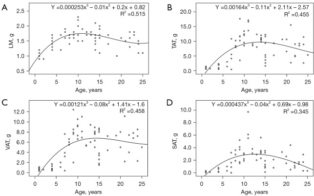
Results: A total of 72 healthy female cynomolgus monkeys, aged 1-25 years, were included in this study. The average lumbar vBMD of female cynomolgus monkeys increased with age until the age of 10 years, around which it reached peak bone mass, with a relatively marked decline after the age of 13 years. The ABLRs of female cynomolgus monkeys in the premenopausal (13-19 years) and postmenopausal age groups (20-25 years) were -4.9% and -21.2%, respectively. Ridge regression analysis showed that age and subcutaneous adipose tissue (SAT) contributed positively to the average lumbar vBMD in animals aged ≤10 years, whereas in animals aged >10 years, age contributed negatively to lumbar vBMD. The RMS-CV% (RMS-SD) of the lumbar vBMD measured using QCT ranged from 0.47% to 1.60% (1.91-6.31 mg/cm3).
Conclusions: Age-related changes in lumbar vBMD measured using QCT in healthy female monkeys showed similar trends to those in humans. Age and SAT may affect the lumbar vBMD in female cynomolgus monkeys. QCT revealed good precision in measuring the lumbar vBMD in female cynomolgus monkeys.
Keywords: Cynomolgus monkeys; precision; quantitative computed tomography (QCT); volumetric bone mineral density (vBMD).
2023 Quantitative Imaging in Medicine and Surgery. All rights reserved.
Conflict of interest statement
Conflicts of Interest: All authors have completed the ICMJE uniform disclosure form (available at https://qims.amegroups.com/article/view/10.21037/qims-22-763/coif). All authors reported that this work was supported by the National Natural Science Foundation of China (No. 81871383). Yuefeng Li is the head of Research and Development Department in Guangdong Landau Biotechnology Co. Ltd. The authors have no other conflicts of interest to declare.
Figures

References
-
- Xiao PL, Cui AY, Hsu CJ, Peng R, Jiang N, Xu XH, Ma YG, Liu D, Lu HD. Global, regional prevalence, and risk factors of osteoporosis according to the World Health Organization diagnostic criteria: a systematic review and meta-analysis. Osteoporos Int 2022;33:2137-53. 10.1007/s00198-022-06454-3 - DOI - PubMed
LinkOut - more resources
Full Text Sources
Research Materials