Review
. 2022 Oct 7:36:27-45.
doi: 10.1016/j.jpra.2022.09.006. eCollection 2023 Jun.

An illustrated anatomical approach to reducing vascular risk during facial soft tissue filler administration - a review

Affiliations
Review

An illustrated anatomical approach to reducing vascular risk during facial soft tissue filler administration - a review

Jeremy Isaac et al. JPRAS Open. .

Abstract

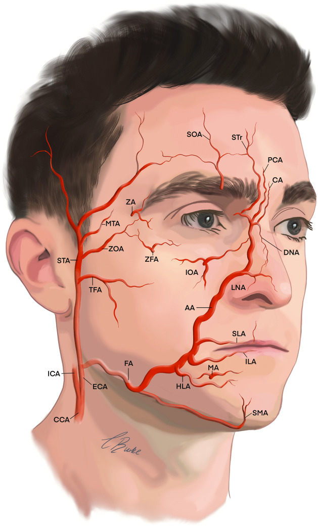
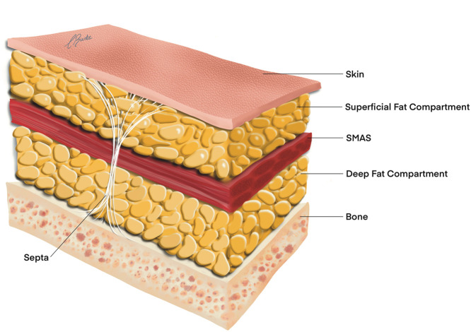
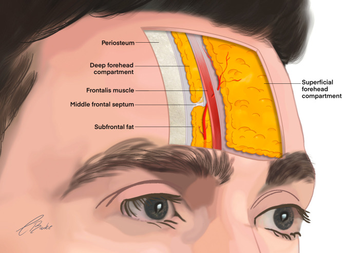
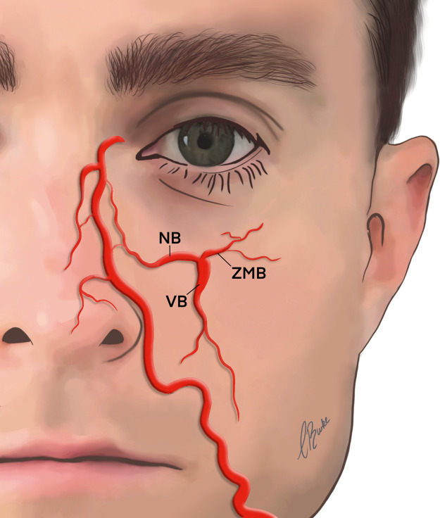
Vascular complications from soft tissue fillers can have catastrophic consequences for patients. Adverse events are rare, but they are increasing, and their appearance may be the result of intravascular injection. A comprehensive understanding of the 2-dimensional anatomy (distribution) and 3-dimensional anatomy (depth) of the facial vasculature is fundamental for the safe delivery of nonsurgical cosmetic procedures. The purpose of this review is to provide an illustrated approach to examine surgical anatomy specific to the facial vascular system and the anatomical considerations clinicians need to give in specific danger during injectable cosmetic procedures. A grounding in safety and anatomy will help the new injector to mitigate the risk of vascular complications.

Keywords: Adverse events; Anatomy; Fillers; Hyaluronic acid (HA).


Figures

Figure 1:
Figure 1
The arterial supply to the face is from the ECA and ICA. Arteries contributing to the vascular supply of the face: AA; angular artery, CA; central artery, CCA; common carotid artery, DNA; dorsal nasal artery, ECA; external carotid artery, FA; facial artery, HLA; horizontal labiomental, ICA; internal carotid artery, ILA; inferior labial artery, IOA; infraorbital artery, LNA; lateral nasal artery, MA; mental artery, MTA; middle temporal artery, PCA; paracentral artery, SLA; superior labial artery, SMA; Submental artery, SOA; supraorbital artery, STA; superficial temporal artery, STr; supratrochlear artery, TFA; transverse facial artery, ZA; zygomaticotemporal artery, ZFA; zygomaticofacial artery, ZOA; zygomatico-orbital artery.
Figure 2:
Figure 2
Five anatomical layers of the face. SMAS; superficial musculoaponeurotic system.
Figure 3:
Figure 3
Sagittal course of the supratrochlear artery in the forehead.
Figure 4:
Figure 4
Branches of the IOA. AA; angular artery, DNA; dorsal nasal artery, IOA; infraorbital artery, NB; nasal artery branch, STA; superficial temporal artery, ZMB; zygomaticomalar artery branch.
Figure 5:
Figure 5
Variations in course and branching pattern of the FA. AA; angular artery, FA; facial artery, DNA; dorsal nasal artery, NB; nasal artery branch, VB; vestibular artery branch, ZMB; zygomaticomalar artery branch. Adapted from Furukawa M, Mathes DW, Anzai Y. Evaluation of the facial artery on computed tomographic angiography using 64-slice multidetector computed tomography: implications for facial reconstruction in plastic surgery. Plast Reconstr Surg 2013; 131: 526–535.
Figure 6:
Figure 6
Variations in coursing patterns of the facial artery. Medial (above left, 42.9%), lateral (above right, 23.2%), and crossing the variations (below left and right, 33.9%). Adapted from Yang H-M, Lee J-G, Hu K-S, et al. New anatomical insights on the course and branching patterns of the facial artery: clinical implications of injectable treatments to the nasolabial fold and nasojugal groove. Plast Reconstr Surg 2014; 133: 1077–1082.
Figure 7:
Figure 7
Variations in branching patterns of the AA. Adapted from Kim YS, Choi DY, Gil YC, Hu KS, Tansatit T, Kim HJ. The anatomical origin and course of the angular artery regarding its clinical implications. Dermatologic Surgery. 2014 Oct 1;40(10):1070-6.
Figure 8:
Figure 8
Relationship of the dorsal nasal artery to procerus and nasalis.
Figure 9:
Figure 9
Classification of the lip into four zones.
Figure 10:
Figure 10
Positional relationship of SLA and ILA.
Figure 11:
Figure 11
Summary of the topographical areas of the face and corresponding risk of vascular complication.
Figure 12:
Figure 12
3-D vasculature areas of the face with vessel signifying the anatomical layer posing the highest risk for intravascular injection. NLF; nasolabial fold.

References

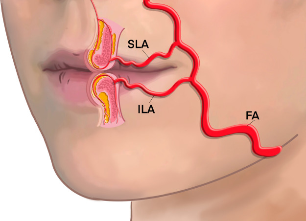
    1. Ghannam S, Sattler S, Frank K, et al. Treating the Lips and Its Anatomical Correlate in Respect to Vascular Compromise. Facial Plast Surg. 2019;35:193–203. - PubMed
    1. Cristel RT, Dayan SH, Akinosun M, et al. Evaluation of Selfies and Filtered Selfies and Effects on First Impressions. Aesthet Surg J. 2021;41:122–130. - PubMed
    1. Heydenrych I, Kapoor KM, De Boulle K, et al. A 10-point plan for avoiding hyaluronic acid dermal filler-related complications during facial aesthetic procedures and algorithms for management. Clin Cosmet Investig Dermatol. 2018;11:603–611. - PMC - PubMed
    1. Schanz S, Schippert W, Ulmer A, et al. Arterial embolization caused by injection of hyaluronic acid (Restylane) Br J Dermatol. 2002;146:928–929. - PubMed
    1. Chatrath V, Banerjee PS, Goodman GJ, et al. Soft-tissue Filler–associated Blindness: A Systematic Review of Case Reports and Case Series. Plast Reconstr Surg Glob Open. 2019;7:e2173. - PMC - PubMed

LinkOut - more resources