Review
. 2023 Mar 5;7(3):216-235.
doi: 10.7150/ntno.81936. eCollection 2023.

Emergence of Raman Spectroscopy as a Probing Tool for Theranostics

Affiliations
Review

Emergence of Raman Spectroscopy as a Probing Tool for Theranostics

Ruchi Singh et al. Nanotheranostics. .

Abstract

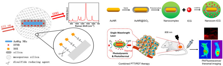
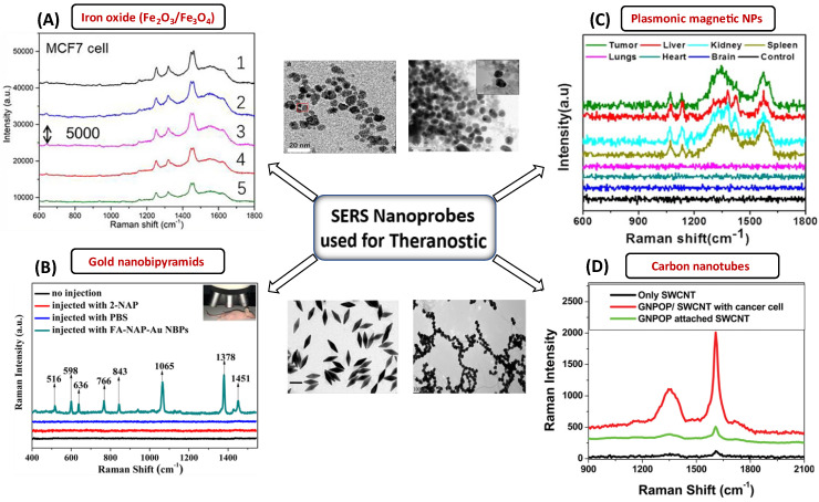
Although medical advances have increased our grasp of the amazing morphological, genetic, and phenotypic diversity of diseases, there are still significant technological barriers to understanding their complex and dynamic character. Specifically, the complexities of the biological systems throw a diverse set of challenges in developing efficient theranostic tools and methodologies that can probe and treat pathologies. Among several emerging theranostic techniques such as photodynamic therapy, photothermal therapy, magnetic resonance imaging, and computed tomography, Raman spectroscopy (RS) is emerging as a promising tool that is a label-free, cost-effective, and non-destructive technique. It can also provide real-time diagnostic information and can employ multimodal probes for detection and therapy. These attributes make it a perfect candidate for the analytical counterpart of the existing theranostic probes. The use of biocompatible nanomaterials for the fabrication of Raman probes provides rich structural information about the biological molecules, cells, and tissues and highly sensitive information down to single-molecule levels when integrated with advanced RS tools. This review discusses the fundamentals of Raman spectroscopic tools such as surface-enhanced Raman spectroscopy and Resonance Raman spectroscopy, their variants, and the associated theranostic applications. Besides the advantages, the current limitations, and future challenges of using RS in disease diagnosis and therapy have also been discussed.

Keywords: Raman and surface-enhanced Raman spectroscopy; Theranostics; nanomedicine; photothermal therapy; plasmonic probes.


Conflict of interest statement

Competing Interests: The authors have declared that no competing interest exists.

Figures

Figure 1
Figure 1
(A) Schematic representation of Raman scattering phenomenon. Figure reprinted from with permission. Copyright (2021) Journal of Applied Physics. (B) Mechanism of enhanced SERS signal of analyte on the noble metal substrate due to electromagnetic and chemical field enhancements. Figure reprinted from with permission. Copyright (2022) The Journal of Physical Chemistry C.
Figure 2
Figure 2
Various probes employed for Raman theranostics such as molecular, plasmonic, carbon, and metal oxide probes. Figure adapted from with permission. Copyright (2019) Journal of Applied Physics, (2015) Frontiers in Chemistry, (2018) Jove-Journal of Visualized Experiments.
Figure 3
Figure 3
Various types of carbon-based probes with different dimensionality and utilization in theranostics applications. Figure reprinted from with permission. Copyright (2019) Chemical Reviews.
Figure 4
Figure 4
(A) Different applications of CNT for cancer diagnosis. Figure reprinted from with permission. Copyright (2021) Journal of Nanobiotechnology (B) Working mechanism of PEG-CNT-ABT737 for treatment of lung cancer cells. Figure reprinted from with permission. Copyright (2021) Journal of Nanobiotechnology. (C) Mechanism of transfer of CNT to blood circulation for treatment. Figure reprinted from with permission. Copyright (2013) Journal of Applied Toxicology. (D) Self-assembly of CNTs into a CNT ring (CNTR) via redox-active poly(4-vinylphenol) (PvPH) brushes. This utilizes a surface-initiated atom transfer radical-polymerization (SI-ATRP) technique to convert Au3+ to Au (0) and encapsulate the CNTR with AuNPs. Figure reprinted from with permission. Copyright (2016) Journal of American Chemical Society.
Figure 5
Figure 5
(A) Applications of plasmonic NPs in biomedical applications. Figure reprinted from with permission. Copyright (2019) Advanced Science. (B) SERS imaging for identification of tumor margin. The red part imaging area shows the increment in the tumor. Figure reprinted from with permission. Copyright (2012) ACS Nano.
Figure 6
Figure 6
(A) Scheme where disulfide bond acting as a nanocarrier in intracellular drug delivery scheme. Figure reprinted from with permission. Copyright (2013) Analytical Chemistry. (B) Fabrication of Nanocom-ICG in combination for PTT/PDT therapy. Figure reprinted from with permission. Copyright (2021) Journal of Nanobiotechnology.
Figure 7
Figure 7
SERS nanomaterials used for the diagnostic purposes (A) Iron-oxide NPs to differentiate between cancerous and normal cells collected from 5 different spots laser in rabbit blood samples. The significant Raman signature bands are at around 1255 cm-1 (C=O stretching) and 1325 cm-1 (CC stretching) observed for targeted tumor cells. Figure reprinted from with permission. Copyright (2022) Fundamental Research. (B) The detection of MCF-7 cancer cells based on SERS properties of conjugated Au NBPs. The prominent bands are observed due to binding of these AuNBPs with MCF-7 cancer cells. Figure reprinted from with permission. Copyright (2017) ACS Biomaterials Science & Engineering. (C) Plasmonic magnetic NPs for intensity profiling of various organs in tumor areas. The spectral intensities at 1330 and 1090 cm-1 are enhanced for tumor targeted areas. Figure reprinted from with permission. Copyright (2021) ACS Applied Bio Materials. D) SERS based cancer cell detection using gold nano-popcorn with SWCNTs. Significant Raman bands at 1300 and 1590 cm-1 that correspond to the (D and G) band of SWCNTs were observed for malignant tissues. Figure reprinted from with permission. Copyright (2011) ACS Applied Materials & Interfaces.

References

    1. Warner S. Diagnostics + therapy = theranostics: strategy requires teamwork, partnering, and tricky regulatory maneuvering. The Scientist. 2004. pp. 38–40.
    1. Canetta E. Current and Future Advancements of Raman Spectroscopy Techniques in Cancer Nanomedicine. International journal of molecular sciences. 2021;22:13141. - PMC - PubMed
    1. Sung H, Ferlay J, Siegel R, Laversanne M, Soerjomataram I, Jemal A. et al. Global cancer statistics 2020: GLOBOCAN estimates of incidence and mortality worldwide for 36 cancers in 185 countries. Ca-a Cancer Journal For Clinicians. 2021;71:209–49. - PubMed
    1. Huang H, Lovell JF. Advanced functional nanomaterials for theranostics. Advanced functional materials. 2017;27:1603524. - PMC - PubMed
    1. Svenson S. Theranostics: Are We There Yet? Molecular Pharmaceutics. 2013;10:848–56. - PubMed

Publication types