Clinical Trial
. 2023 Apr 6:2023:9412132.
doi: 10.1155/2023/9412132. eCollection 2023.

Self-Expanding Versus Balloon Expanding Coronary Stents in Intervention of the Degenerated Saphenous Vein Graft: Memmingen Coronary Artery Bypass Stenosis Trial (MECAST)

Affiliations
Clinical Trial

Self-Expanding Versus Balloon Expanding Coronary Stents in Intervention of the Degenerated Saphenous Vein Graft: Memmingen Coronary Artery Bypass Stenosis Trial (MECAST)

Marcus Siry et al. J Interv Cardiol. .

Abstract

Objectives: The aim of this retrospective analysis was to compare the patient outcome after interventional therapy of saphenous vein graft (SVG) stenoses in an all-comers population receiving either self-expanding drug-eluting stents (SExS) or balloon expanding drug-eluting stents (BExS).

Background: The interventional therapy of degenerated SVGs remains challenging. Diameter variations of stenotic segments and friable plaques can lead to malapposition and distal embolization with increased major adverse cardiac event (MACE) rates.

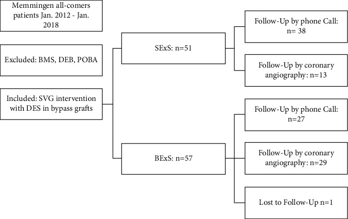
Methods: 107 patients with a total of 130 SVG interventions were separated into two groups according to either SExS (n = 51) or BExS (n = 56) treatment. Primary endpoint was the MACE rate, which is defined as the composite of cardiac death, myocardial infarction (MI), target vessel (TVR), and target lesion revascularization (TLR) at 30 days and at one-year follow-up.

Results: Both patient groups did not differ significantly regarding patient characteristics. The patient outcome was significantly better in the SExS patient group: the MACE rate at 30 days was 1/51 (2.0%) in group SExS vs. 7/56 (12.5%) in group BExS; p < 0.05. At one-year follow-up, the MACE rate remained significantly lower in the SExS group 8/51(15.7%) vs. 20/56 (35.7%) in the BExS group, p < 0.02. Additionally, cardiac death occurred significantly later within the SExS patient group compared to the BExS group (p < 0.05). A better overall outcome of patients with de novo SVG-stenosis compared to patients with previous CABG (coronary artery bypass graft) intervention was noted in both groups.

Conclusion: Our findings demonstrate that SVG treatment with SExS is safe and provides clinical benefits by comparatively improving short and especially long-term patient outcomes.


Conflict of interest statement

The authors declare that they have no conflicts of interest.

Figures

Figure 1
Figure 1
Delivery of self-expanding drug-eluting stent. (a) Stent is mounted on a semi-compliant balloon and is restrained by a splittable sheath. (b) Balloon inflation splits the sheath and releases the self-expanding stent. (c) Balloon is then deflated, leaving the 0.0032″ sheath between the stent and the vessel wall. (d) Balloon and sheath are then withdrawn leaving the stent opposed to the vessel wall. The two radiopaque stent markers are located at the edges of the stent (red arrows). With permission by STENTYS.
Figure 2
Figure 2
(a) SVG of RCA with stenosis located in proximal shaft: (b) modified floating wire technique. (a) SVG-stenosis located in proximal shaft, SExS (Xposition S® 3.0–3.5 ∗ 27 mm) implantation (A), sheath retrieval (B) with support of coronary wire (C). (b) Proximal SVG-RCA stenosis with implantation of Xposition S 3.0–3.5 ∗ 27 mm, sheath retrieval with insertion of an additional wire through the guiding catheter floating free in the ascending aorta => prevention of deep intubation with possible subsequent distal wire perforation. RCA = right coronary artery; SVG = saphenous vein graft; SExS: self-expanding drug eluting stent.
Figure 3
Figure 3
Retrospective data analysis. SVG = saphenous vein graft; SExS = self-expanding drug eluting stent; BExS = balloon expanding drug eluting stent; BMS = bare metal stent; DEB = drug eluting balloon; and POBA = plane old balloon angioplasty.
Figure 4
Figure 4
(a) MACE timeline of SExS and BExS patient groups. MACE = major adverse cardiac event; SExS = self-expanding drug eluting stent; BExS = balloon expanding drug eluting stent; TVR = target vessel revascularization; TLR = target lesion revascularization; AMI = acute myocardial infarction. (b) Mean event rates in BExS patient group compared to SExS group. SExS = self-expanding drug eluting stent; BExS = balloon expanding drug eluting stent; MACE = major adverse cardiac event; AMI = acute myocardial infarction; TLR = target lesion revascularization; TVR = target vessel revascularization.
Figure 5
Figure 5
(a) Kaplan–Meier curve for cumulative MACE-free survival within 30 days in BExS and SExS patient groups (log rank p < 0.05). SExS = self-expanding drug eluting stent; BExS = balloon expanding drug eluting stent. (b) Central illustration: Kaplan–Meier curve for cumulative MACE-free survival within one year in BExS and SExS patient groups (log rank p < 0.02). SExS = self-expanding drug eluting stent; BExS = balloon expanding drug eluting stent. (c) Kaplan–Meier curve for cardiac death within 12 months in the BExS and SExS patient groups (log rank p < 0.05). SExS = self-expanding drug eluting stent; BExS = balloon expanding drug eluting stent.
Figure 6
Figure 6
Forest plot depicting hazard ratios of several covariates. Statistical significance (p < 0.01) marked with a star. A hazard ratio <1 illustrates a protective effect on patient outcome, whereas a hazard ratio >1 represents a negative effect. ACS = acute coronary syndrome; EF = ejection fraction; PAOD = peripheral artery occlusive disease, EPD = embolic protection device; CABG = coronary artery bypass graft; TIMI = thrombolysis in myocardial infarction; and SVG = saphenous vein graft.

References

    1. ElBardissi A. W., Aranki S. F., Sheng S., O’Brien S. M., Greenberg C. C., Gammie J. S. Trends in isolated coronary artery bypass grafting: an analysis of the society of thoracic surgeons adult cardiac surgery database. The Journal of Thoracic and Cardiovascular Surgery . 2012;143(2):273–281. doi: 10.1016/j.jtcvs.2011.10.029. - DOI - PubMed
    1. Neumann F.-J., Hochholzer W., Siepe M. ESC/EACTS-leitlinien zur myokardrevaskularisation 2018. Herz . 2018;43(8):689–694. doi: 10.1007/s00059-018-4764-5. - DOI - PubMed
    1. Goldman S., Zadina K., Moritz T., et al. Long-term patency of saphenous vein and left internal mammary artery grafts after coronary artery bypass surgery. Journal of the American College of Cardiology . 2004;44(11):2149–2156. doi: 10.1016/j.jacc.2004.08.064. - DOI - PubMed
    1. Kalan J. M., Roberts W. C. Morphologic findings in saphenous veins used as coronary arterial bypass conduits for longer than 1 year: necropsy analysis of 53 patients, 123 saphenous veins, and 1865 five-millimeter segments of veins. American Heart Journal . 1990;119(5):1164–1184. doi: 10.1016/s0002-8703(05)80249-2. - DOI - PubMed
    1. Baim D. S., Wahr D., George B., et al. Randomized trial of a distal embolic protection device during percutaneous intervention of saphenous vein aorto-coronary bypass grafts. Circulation . 2002;105(11):1285–1290. doi: 10.1161/01.cir.0000012783.63093.0c. - DOI - PubMed

Publication types

LinkOut - more resources