. 2023 Apr 6:2023:7707452.
doi: 10.1155/2023/7707452. eCollection 2023.

Bioaccumulation of CdSe Quantum Dots Show Biochemical and Oxidative Damage in Wistar Rats

Affiliations

Bioaccumulation of CdSe Quantum Dots Show Biochemical and Oxidative Damage in Wistar Rats

Kishan Das et al. Oxid Med Cell Longev. .

Abstract

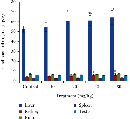
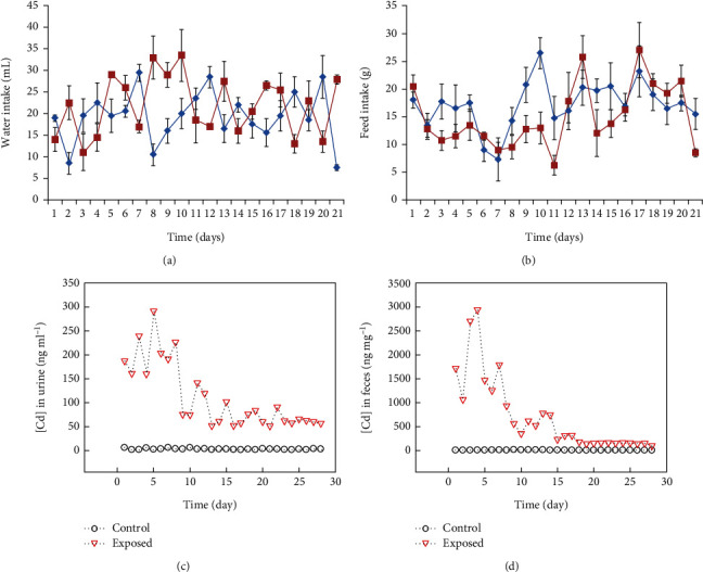
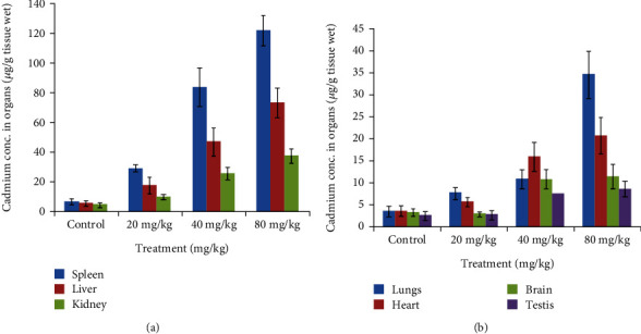
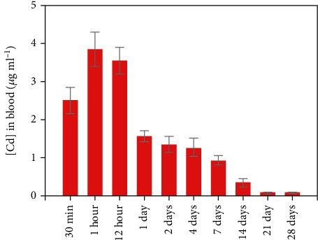
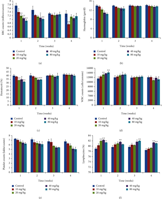
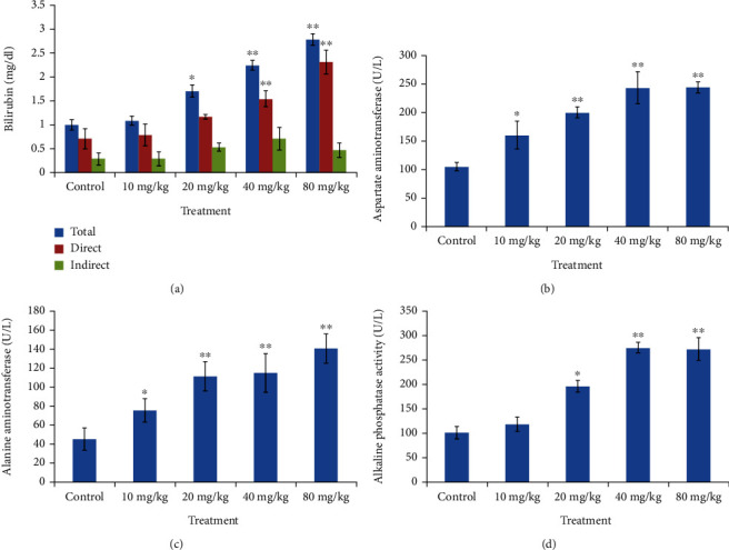
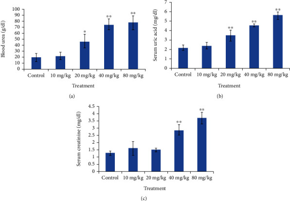
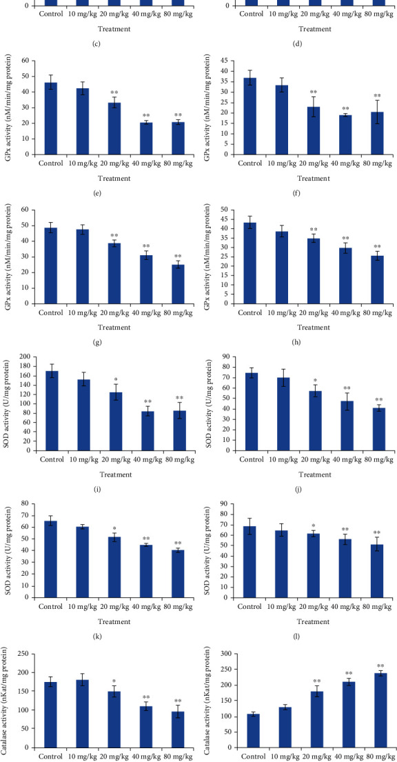
Cadmium selenium quantum dots (CdSe QDs) with modified surfaces exhibit superior dispersion stability and high fluorescence yield, making them desirable biological probes. The knowledge of cellular and biochemical toxicity has been lacking, and there is little information on the correlation between in vitro and in vivo data. The current study was carried out to assess the toxicity of CdSe QDs after intravenous injection in Wistar male rats (230 g). The rats were given a single dose of QDs of 10, 20, 40, and 80 mg/kg and were kept for 30 days. Following that, various biochemical assays, hematological parameters, and bioaccumulation studies were carried out. Functional as well as clinically significant changes were observed. There was a significant increase in WBC while the RBC decreased. This suggested that CdSe quantum dots had inflammatory effects on the treated rats. The various biochemical assays clearly showed that high dose induced hepatic injury. At a dose of 80 mg/kg, bioaccumulation studies revealed that the spleen (120 g/g), liver (78 g/g), and lungs (38 g/g) accumulated the most. In treated Wistar rats, the bioretention profile of QDs was in the following order: the spleen, liver, kidney, lungs, heart, brain, and testis. The accumulation of these QDs induced the generation of intracellular reactive oxygen species, resulting in an alteration in antioxidant activity. It is concluded that these QDs caused oxidative stress, which harmed cellular functions and, under certain conditions, caused partial brain, kidney, spleen, and liver dysfunction. This is one of the most comprehensive in vivo studies on the nanotoxicity of CdSe quantum dots.


Conflict of interest statement

The authors declare that they have no conflicts of interest.

Figures

Figure 1
Figure 1
Organ coefficient (the liver, kidney, testis, spleen, and brain) of control and treated set of Wistar rats. Organ coefficient is the ratio of weight of organs (mg) to weight of animals (g). Statistically significant results at p < 0.05.
Figure 2
Figure 2
Metabolic response of the Wistar rats (a) depicts water intake and (b) depicts food intake, with block lines representing the control and red line treated groups, respectively, graph showing the concentration of Cd in (c) urine and (d) feces in the control and exposed groups of rats.
Figure 3
Figure 3
Graph depicting bioaccumulation of cadmium in different organs, with the spleen showing the maximum accumulation and testis the least.
Figure 4
Figure 4
Graph showing the concentration of Cd in blood of Wistar rats administered with CdSe QDs.
Figure 5
Figure 5
Hematological results from animals treated with varying dose of CdSe QDs. (a) Red blood cells (millions/cumm). (b) Hemoglobin (g/dL). (c) Hematocrit (%). (d) White blood cells (1000 cells/cumm). (e) Platelet counts (lakhs/cumm). (f) Lymphocytes (%). The results show the mean and standard deviation of six independent readings.
Figure 6
Figure 6
Graph depicting changes in liver function markers from Wistar rats treated with varying dose of CdSe QDs. (a) Bilirubin (total, direct, and indirect). (b) Asparate aminotransferase (AST). (c) Alanine aminotransferase (ALT). (d) Alkaline phosphatase (ALP). Statistically significant at (p < 0.05), ∗∗very significant (p < 0.001).
Figure 7
Figure 7
Graph depicting dose dependent changes in kidney function markers in the treated Wistar rates. (a) Blood urea. (b) Uric acid. (c) Creatinine. Significant (p < 0.05) and ∗∗very significant changes (p < 0.001).
Figure 8
Figure 8
Graph depicting comparison of antioxidative enzyme (the liver, kidney, and brain, respectively) in the control and CdSe QDs-treated Wistar rats. Lipid peroxidise activity in the (a) liver, (b) kidney, (c) brain, and (d) spleen. GPx activity in the (e) liver, (f) kidney, (g) brain, and (h) spleen. SOD activity in the (i) liver, (j) kidney, (k) brain, and (l) spleen. Catalase activity in the (m) liver, (n) kidney, (o) brain, and (p) spleen.

References

    1. Caruthers S. D., Wickline S. A., Lanza G. M. Nanotechnological applications in medicine. Current Opinion in Biotechnology . 2007;18(1):26–30. doi: 10.1016/j.copbio.2007.01.006. - DOI - PubMed
    1. Thomas T. Nanotechnology in consumer products: addressing potential health and ssafety implications for consumers. In: Monteiro-Riviere N. A., Tran C. L., editors. Nanotoxicology Progress toward Nanomedicine . Boca Raton, Boca Raton: CRC Press; 2014. pp. 97–112. - DOI
    1. Joksimovic N., Selakovic D., Jovicic N., et al. Nanoplastics as an invisible threat to humans and the environment. Journal of Nanomaterials . 2022;2022(12):p. 15. doi: 10.1155/2022/6707819.6707819 - DOI
    1. Qian X., Peng X.-H., Ansari D. O., et al. In vivo tumor targeting and spectroscopic detection with surface-enhanced Raman nanoparticle tags. Nature Biotechnology . 2008;26(1):83–90. doi: 10.1038/nbt1377. - DOI - PubMed
    1. McMahon S. J., Hyland W. B., Muir M. F., et al. Biological consequences of nanoscale energy deposition near irradiated heavy atom nanoparticles. Scientific Reports . 2011;1(1):p. 18. doi: 10.1038/srep00018. - DOI - PMC - PubMed

LinkOut - more resources