. 2023 Mar 27;13(6):2015-2039.
doi: 10.7150/thno.80615. eCollection 2023.

Recent advances in GelMA hydrogel transplantation for musculoskeletal disorders and related disease treatment

Affiliations

Recent advances in GelMA hydrogel transplantation for musculoskeletal disorders and related disease treatment

Bin Lv et al. Theranostics. .

Abstract

Increasing data reveals that gelatin that has been methacrylated is involved in a variety of physiologic processes that are important for therapeutic interventions. Gelatin methacryloyl (GelMA) hydrogel is a highly attractive hydrogels-based bioink because of its good biocompatibility, low cost, and photo-cross-linking structure that is useful for cell survivability and cell monitoring. Methacrylated gelatin (GelMA) has established itself as a typical hydrogel composition with extensive biomedical applications. Recent advances in GelMA have focused on integrating them with bioactive and functional nanomaterials, with the goal of improving GelMA's physical, chemical, and biological properties. GelMA's ability to modify characteristics due to the synthesis technique also makes it a good choice for soft and hard tissues. GelMA has been established to become an independent or supplementary technology for musculoskeletal problems. Here, we systematically review mechanism-of-action, therapeutic uses, and challenges and future direction of GelMA in musculoskeletal disorders. We give an overview of GelMA nanocomposite for different applications in musculoskeletal disorders, such as osteoarthritis, intervertebral disc degeneration, bone regeneration, tendon disorders and so on.

Keywords: bone; hydrogel; musculoskeletal disorders; nanomaterials; tissue engineering.


Conflict of interest statement

Competing interests: Overcoming PARP inhibitor resistance through the combination of RTK inhibi‑ tors and PARPi, are covered in the provisional patent UTSC.P1450US.P1.

Figures
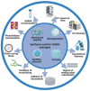
Figure 1
Figure 1
Schematic illustration of the synthesis and photocrosslinking of hydrogel in this review. GelMA hydrogel was modified to generate desired physical or chemical properties.
Figure 2
Figure 2
Systematic diagram illustrates the main content of this review. Applications of GelMA-based hydrogel systems for musculoskeletal disorders.
Figure 3
Figure 3
A) Preparation method of GelMA hydrogel. B) Different application forms of GelMA-based hydrogel systems for musculoskeletal disorders.
Figure 4
Figure 4
Engineering GelMA hydrogel for cartilage tissue. A) Schematic overview of the GelMA@eIm/ZIF-67 hydrogel for Co-ion release to enhance osteogenesis and angiogenesis through activating HIF-1α. B) Synthesis and digital images of the GelMA and eIm/ZIF-67-based hydrogels. C) Assessment of osteogenic differentiation. D) Micro-CT images of bone tissue. E) Expression level of VEGF and HIF-1α. Reproduced and adapted with permission from ref. . Copyright 2021 American Chemical Society.
Figure 5
Figure 5
GelMA-loaded nanoplatelets in bone tissue engineering. A) Schematic overview of GelMA bioprinting of the scaffolds for osteochondral repair. Reproduced with permission from ref . Copyright 2019 Wiley. B) GelMA scaffold preparation by photocuring GelMA bioprinting and lyophilization. C) Structure properties and biocompatibility of GelMA scaffolds. Reproduced with permission from ref. . Copyright 2018 American Chemical Society. D) Gross images and Micro-CT observation of osteochondral repair by scaffolds. Reproduced with permission from . Copyright 2021 Elsevier.
Figure 6
Figure 6
GelMA-based hydrogel for intervertebral disc engineering. A) Schematic overview of GelMA scaffolds for intervertebral disc degeneration. B) Expression of Col II of NPCs-loaded hydrogel. Reproduced and adapted with permission from ref . Copyright 2021 Elsevier. C) GelMA hydrogel for preventing recurrence after partial discectomy. Reproduced and adapted with permission from ref . Copyright 2020 Wiley. D) Expression level of aggrecan of NPCs-loaded hydrogel. Reproduced and adapted with permission from ref . Copyright 2018 Elsevier.
Figure 7
Figure 7
Implantation of GelMA-based hydrogel for tendon disorders. A) The fabrication process of the siRNA@MS@HA hydrogel. Reproduced and adapted with permission from ref . Copyright 2022 Wiley. B) Timeline and diagram of achilles tendon repair. Reproduced and adapted with permission from ref . Copyright 2022 Wiley. C) Schematic diagram of macroscopic images in the antiadhesion barrier in peritendinous tissue. Reproduced and adapted with permission from ref . Copyright 2022 Wiley. D) Immunofluorescence images of expression level of tendon tissues. Reproduced and adapted with permission from ref . Copyright 2022 Wiley.
Figure 8
Figure 8
GelMA-loaded hydrogel for skeletal muscle regeneration. A) Schematic overview of fabrication of micropatterned and unpatterned GelMA fibers. B) FE-SEM photographs show myoblasts on GelMA fibers. C) Fluorescent photographs of myotubes on GelMA fibers. Reproduced and adapted with permission from ref . Copyright 2018 Wiley. D) Fluorescent photographs of AChR clusters on myotubes. E) Biocompatibility and conductivity of GelMA-based MXene and AuNPs hydrogels. Reproduced and adapted with permission from and ref . Copyright 2021 American Chemical Society.
Figure 9
Figure 9
GelMA-DA hydrogel in neural tissue engineering. A) Illustration of cell-laden bioprinting by GelMA-based bioinks. B) Printability evaluation using GelMA-based bioinks. C) Cell-laden bioprinting by GelMA-based bioink. D) Optical and surface plotting photographs of photo-patterned hydrogels. E) Inputting AutoCAD patterns and the patterns by photocurable GelMA-based hydrogels. F) Immunofluorescence photographs of DRG cells-loaded GelMA hydrogel. Reproduced and adapted with permission from ref . Copyright 2019 Elsevier.
Figure 10
Figure 10
Schematic overview of the conductive NGC in peripheral nerve. A) Schematic illustration of NGC. B) Modification and characterization of the rGO/BDNF/GelMA. C) Construction and characterization of the NGCs. D) Influence of rGO/BDNF/GelMA on NSCs growth and proliferation. E) Influence of rGO/BDNF/GelMA on NSC differentiation and neurite extension. Reproduced and adapted with permission from ref . Copyright 2022 American Chemical Society.
Figure 11
Figure 11
MXene-based GelMA hydrogel with osteogenicity for osteosarcoma. A) Schematic overview of multifunctional implants after osteosarcoma excision. B) Illustration of process for preparation of GelMA-based multifunctional substrate. C) Infrared thermal images of GelMA-based hydrogel. D) SEM images of osteoblasts cultured on multifunctional substrates. E) Histological assessment of bone contact. Reproduced and adapted with permission from ref . Copyright 2020 American Chemical Society.
Figure 12
Figure 12
Molecular mechanism of GelMA-based materials for musculoskeletal diseases. A) GelMA-PPy-Fe promotes bone regeneration by regulating the NOTCH/MAPK/SMAD pathway and the Wnt/β-Catenin pathway. B) GelMA-g-GSH promotes bone regeneration through the PI3K/Akt signaling pathway. C) MPEG/PCL/GelMA mechanism for the treatment of PNI. D) Mechanism of PEGDA/GelMA in the treatment of bone tumors.

References

    1. Cieza A, Causey K, Kamenov K, Hanson SW, Chatterji S, Vos T. Global estimates of the need for rehabilitation based on the Global Burden of Disease study 2019: a systematic analysis for the Global Burden of Disease Study 2019. Lancet. 2021;396:2006–17. - PMC - PubMed
    1. Charalambides C, Beer M, Cobb AG. Poor results after augmenting autograft with xenograft (Surgibone) in hip revision surgery: a report of 27 cases. Acta Orthop. 2005;76:544–9. - PubMed
    1. Dong Z, Yuan Q, Huang K, Xu W, Liu G, Gu Z. Gelatin methacryloyl (GelMA)-based biomaterials for bone regeneration. RSC Adv. 2019;9:17737–44. - PMC - PubMed
    1. Gong T, Xie J, Liao J, Zhang T, Lin S, Lin Y. Nanomaterials and bone regeneration. Bone Res. 2015;3:15029. - PMC - PubMed
    1. Li G, Che MT, Zhang K, Qin LN, Zhang YT, Chen RQ. et al. Graft of the NT-3 persistent delivery gelatin sponge scaffold promotes axon regeneration, attenuates inflammation, and induces cell migration in rat and canine with spinal cord injury. Biomaterials. 2016;83:233–48. - PubMed

Publication types