Review
. 2023 Mar 29:14:1145233.
doi: 10.3389/fphys.2023.1145233. eCollection 2023.

Vitamin D and chronic kidney disease: Insights on lipid metabolism of tubular epithelial cell and macrophages in tubulointerstitial fibrosis

Affiliations
Review

Vitamin D and chronic kidney disease: Insights on lipid metabolism of tubular epithelial cell and macrophages in tubulointerstitial fibrosis

Luís Eduardo D Gonçalves et al. Front Physiol. .

Abstract

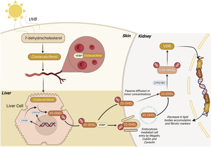
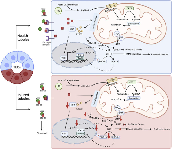
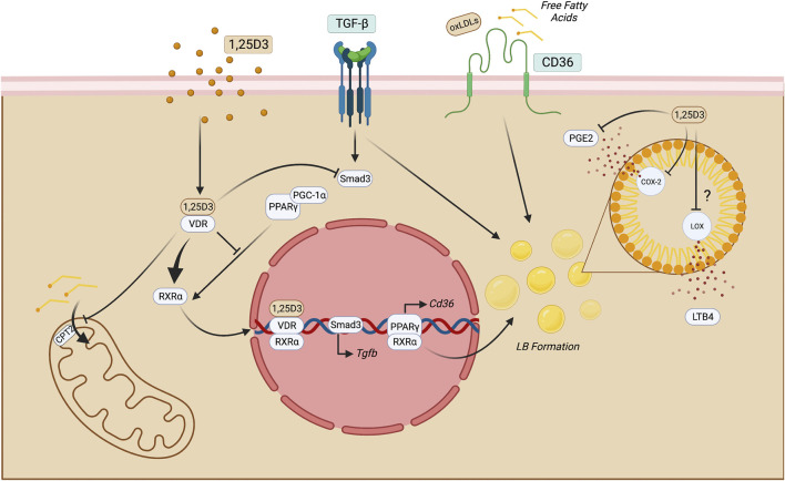
Chronic kidney disease (CKD) has been recognized as a significant global health problem due to being an important contributor to morbidity and mortality. Inflammation is the critical event that leads to CKD development orchestrated by a complex interaction between renal parenchyma and immune cells. Particularly, the crosstalk between tubular epithelial cells (TECs) and macrophages is an example of the critical cell communication in the kidney that drives kidney fibrosis, a pathological feature in CKD. Metabolism dysregulation of TECs and macrophages can be a bridge that connects inflammation and fibrogenesis. Currently, some evidence has reported how cellular lipid disturbances can affect kidney disease and cause tubulointerstitial fibrosis highlighting the importance of investigating potential molecules that can restore metabolic parameters. Vitamin D (VitD) is a hormone naturally produced by mammalian cells in a coordinated manner by the skin, liver, and kidneys. VitD deficiency or insufficiency is prevalent in patients with CKD, and serum levels of VitD are inversely correlated with the degree of kidney inflammation and renal function. Proximal TECs and macrophages produce the active form of VitD, and both express the VitD receptor (VDR) that evidence the importance of this nutrient in regulating their functions. However, whether VitD signaling drives physiological and metabolism improvement of TECs and macrophages during kidney injury is an open issue to be debated. In this review, we brought to light VitD as an important metabolic modulator of lipid metabolism in TECs and macrophages. New scientific approaches targeting VitD e VDR signaling at the cellular metabolic level can provide a better comprehension of its role in renal physiology and CKD progression.

Keywords: VDR (vitamin D receptor); beta-oxidation; immune cell; metabolism; renal fibrosis.


Conflict of interest statement

The authors declare that the research was conducted in the absence of any commercial or financial relationships that could be construed as a potential conflict of interest.

Figures

FIGURE 1
FIGURE 1
Metabolic route of 1,25 dihydroxyvitamin D3 synthesis. Firstly, 7-dehydrocholesterol (7-DHC) is converted to pre-vitamin D3 (Cholecalciferol) through the action of UVB light in the skin. Subsequently, this compound migrates to the bloodstream where it binds to VDBP, and the complex is transported to the liver. Then, the liver cell promotes the conversion of pre-vitamin D3 into 25-hydroxyvitamin D3 (25-OHD3) mediated by CYP27A1 and CYP2R1. Finally, 25-OHD3 migrates to the kidney where it can be passively diffused to the cells or be endocytosed via megalin, cubilin and caveolin membrane receptors. Once 25-OHD3 reaches the cytosol, it is converted by CYP27B1 into 1,25 dihydroxyvitamin D3 (1,25D3), the bioactive form of VitD. Subsequently, the 1,25D3 binds to VDR, migrate to the nucleus, and heterodimerizes with other nuclear hormones receptors, leading to the transcription of genes related to lipid metabolism. 7-DHC, 7-dehydrocholesterol; UVB, ultraviolet B (radiation); VDBP, vitamin D binding protein; VitD, vitamin D. Figure created with BioRender.com.
FIGURE 2
FIGURE 2
Proposal mechanism of VitD on healthy and injured tubular cell metabolism. FAO (or β-oxidation) is the main energy source of TECs. In the cytoplasm, FAs are transformed into Acyl-CoA by the enzyme Acyl-CoA synthetase. Then, the mitochondrial enzyme CPT1a converts acyl-CoA in acylcarnitine, which can be transported into mitochondrial matrix via carnitine/acylcarnitine translocase. Subsequently, acylcarnitine is subjected to the CPT2 activity to be reconverted into acyl-CoA and the FAO is initiated: acyl-CoA is converted into acetyl-CoA, which in turn, fuels the TCA cycle that provides reducing equivalents to ETC to produce ATP. VitD may be an important regulator of the FAO in TECs. After internalization via passive diffusion or receptor-mediated endocytosis, 25-hydroxyvitamin D3 is converted into its bioactive form, 1,25D3. The recognition of the active form by VDR causes transcriptional changes and leads to SIRT1 expression, which in turn, promotes the inhibition of profibrotic pathways, such as SMAD, and contributes to the deacetylation of PGC1 α and, thus, sustaining the FAO. In addition, VitD can also activate SIRT1 via AMPK activation, which increases NAD+ production. In the injured tubules the levels of endocytic receptors are reduced, impairing the optimal internalization of 25-OHD3 (calcitriol), and compromising the VitD activity. The reduced levels of VitD decrease the SIRT1 and SIRT3 expression as well as diminish the function of both PCG1 α and CPT1a. These alterations disrupt the FAO and contribute to profibrotic responses. 1,25D3, 1,25 dihydroxyvitamin D3; 25-OHD3, 25-hydroxyvitamin D3; AMPK, adenosine monophosphate-activated protein kinase; CPT, carnitine palmitoyltransferase; FA, fatty acids; FAO, fatty acid oxidation; PGC-1α, peroxisome proliferator-activated receptor-gamma coactivator-1α; TCA, tricarboxylic acid cycle; TECs, tubular epithelial cells; VDR, vitamin D receptor; VDBP, vitamin D binding protein. ETC, electron transport chain. Figure created with BioRender.com.
FIGURE 3
FIGURE 3
Putative mechanisms of 1,25D3 function in macrophages. Once 1,25D3 binds to VDR, it heterodimerizes with RXRα and translocate to the nucleus, impairing PPARγ-mediated functions. This may impact LB formation and, therefore, reduce the production of eicosanoids, such as LTB4 and PGE2. The latter may also be reduced through direct inhibition of COX-2 through 1,25D3. PPARγ blockade also may reduce CD36 exposure on the cell surface, leading to decreased lipid uptake. In macrophages, this process may limit FAO by inhibiting CPT2 and consequent long-chain fatty acid transport into the mitochondria. Moreover, since the fibrotic milieu is enriched with TGF-β, its signaling triggers Smad-3 and lipid accumulation in macrophages suggesting that this cytokine may have roles beyond fibrosis induction. 1,25 dihydroxyvitamin D3; COX-2, cicloxygenase-2; LOX, lipoxygenase; CPT, carnitine palmitoyltransferase; FAO, fatty acid oxidation; LB, lipid bodies; LTB4, leukotriene B4; PGE2, Prostaglandin E2; TECs, tubular epithelial cells; VDR, vitamin D receptor. Figure created with BioRender.com.

Similar articles

Cited by

References

    1. Barsony J., Prufer K. (2002). Vitamin D receptor and retinoid X receptor interactions in motion. Vitam. Horm. 65, 345–376. 10.1016/s0083-6729(02)65071-x - DOI - PubMed
    1. Batista-Gonzalez A., Vidal R., Criollo A., Carreno L. J. (2019). New insights on the role of lipid metabolism in the metabolic reprogramming of macrophages. Front. Immunol. 10, 2993. 10.3389/fimmu.2019.02993 - DOI - PMC - PubMed
    1. Bhargava P., Schnellmann R. G. (2017). Mitochondrial energetics in the kidney. Nat. Rev. Nephrol. 13, 629–646. 10.1038/nrneph.2017.107 - DOI - PMC - PubMed
    1. Bhatia D., Chung K. P., Nakahira K., Patino E., Rice M. C., Torres L. K., et al. (2019). Mitophagy-dependent macrophage reprogramming protects against kidney fibrosis. JCI Insight 4, e132826. 10.1172/jci.insight.132826 - DOI - PMC - PubMed
    1. Bikle D., Christakos S. (2020). New aspects of vitamin D metabolism and action - addressing the skin as source and target. Nat. Rev. Endocrinol. 16, 234–252. 10.1038/s41574-019-0312-5 - DOI - PubMed

LinkOut - more resources