Review
. 2023 Mar 31:14:1154374.
doi: 10.3389/fphys.2023.1154374. eCollection 2023.

Inhibition of the calcium-sensing receptor by extracellular phosphate ions and by intracellular phosphorylation

Affiliations
Review

Inhibition of the calcium-sensing receptor by extracellular phosphate ions and by intracellular phosphorylation

Patricia P Centeno et al. Front Physiol. .

Abstract

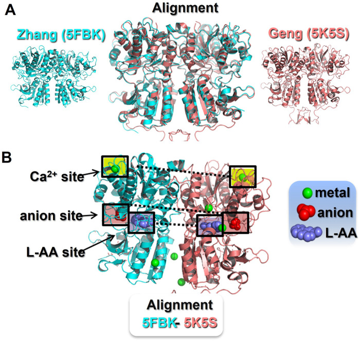
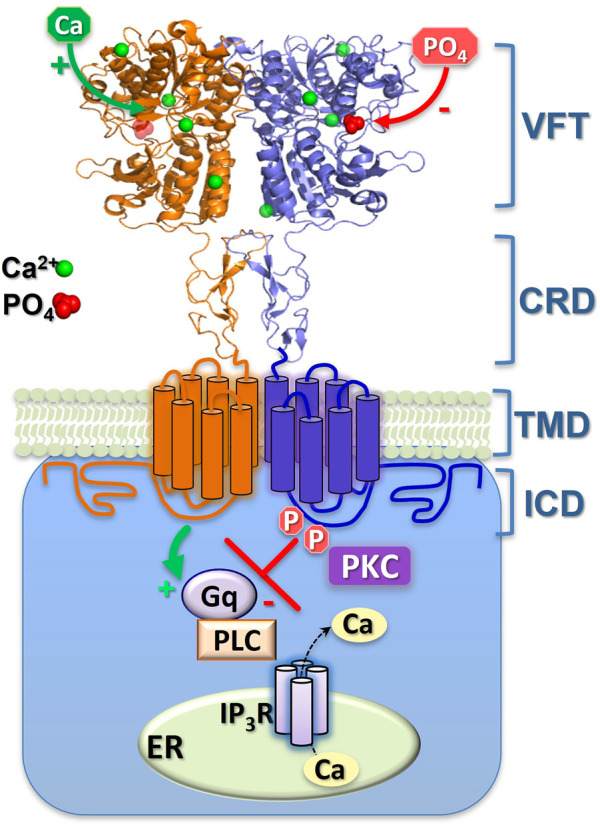
As both a sensor of extracellular calcium (Ca2+ o) concentration and a key controller of Ca2+ o homeostasis, one of the most interesting properties of the calcium-sensing receptor (CaR) is its sensitivity to, and modulation by, ions and small ligands other than Ca2+. There is emerging evidence that extracellular phosphate can act as a partial, non-competitive CaR antagonist to modulate parathyroid hormone (PTH) secretion, thus permitting the CaR to integrate mineral homeostasis more broadly. Interestingly, phosphorylation of certain intracellular CaR residues can also inhibit CaR responsiveness. Thus, negatively charged phosphate can decrease CaR activity both extracellularly (via association with arginine) and intracellularly (via covalent phosphorylation).

Keywords: calcium-sensing receptor; parathyroid hormone; phosphate-sensing; receptor phosphorylation; secondary hyperparathyroidism.


Conflict of interest statement

The authors declare that the research was conducted in the absence of any commercial or financial relationships that could be construed as a potential conflict of interest.

Figures

FIGURE 1
FIGURE 1
Identification of ionic binding sites in the CaR extracellular domain. Panel (A) Sequence alignment and structural superposition of the currently available CaR ECD models in the active conformation. The Zhang et al. (2016) model (5FBK) is shown in blue (left) and the Geng et al. (2016) model (5K5S) is shown in pink (right). In the middle, an alignment of both models reveals an almost identical CaR ECD structure with minor differences. Pymol root mean square deviation (RMSD) 0.5, after 5 cycles of iteration. Panel (B) Active conformation of the CaR ECD with reported ligand binding sites. The Zhang model (5FBH) is shown as the left monomer, with the Geng model (5K5S) on the right. The Zhang model describes three Ca2+-binding sites, one in the upper domain and two in the lower domain facing the interface between monomers. At the same locations, the Geng model describes two Ca2+-binding sites, but two additional Ca2+-binding sites in the cleft between the upper and lower domains. Both models identified a common L-amino acid binding site and a common anion binding site, both located at the cleft between the upper and lower domains. In addition, the Geng model includes an anion binding site located in the lower domain. The ligand binding sites highlighted in boxes are those found in both crystal models. L-AA, L-amino acid.
FIGURE 2
FIGURE 2
Schematic representation of CaR inhibition by extracellular phosphate ions and by intracellular, covalent phosphorylation. The binding-sites of the (activating) Ca2+ and (inhibitory) phosphate (PO4) ions shown here are approximate, though their 4:1 ratio is consistent with both the crystal models and functional Hill coefficients. Sustained phosphorylation (P) of ICD residues CaRS875 and CaRT888 supresses Ca2+ i mobilisation. Episodic dephosphorylation of CaRT888 (at least) permits Ca2+ i oscillations, while continuous dephosphorylation of these sites elicits enhanced, sustained Ca2+ i mobilisation. The two monomers of the CaR homodimer are shown either as orange or blue. Gq, G protein-q/11; PLC, phospholipase C; IP3R, IP3 receptor; ER, Ca2+ stores of the endoplasmic reticulum; PKC, protein kinase C. The CaR domains shown are the venus flytrap (VFT), cysteine-rich domain (CRD), transmembrane domain (TMD) and intracellular domain (ICD).

Similar articles

Cited by

References

    1. Agoro R., White K. E. (2023). Regulation of FGF23 production and phosphate metabolism by bone-kidney interactions. Nat. Rev. Nephrol. Jan. 9, 185–193. 10.1038/s41581-022-00665-x - DOI - PubMed
    1. Almaden Y., Canalejo A., Hernandez A., Ballesteros E., Garcia-Navarro S., Torres A., et al. (1996). Direct effect of phosphorus on PTH secretion from whole rat parathyroid glands in vitro . J. Bone Min. Res. 11, 970–976. 10.1002/jbmr.5650110714 - DOI - PubMed
    1. Almaden Y., Felsenfeld A. J., Rodriguez M., Canadillas S., Luque F., Bas A., et al. (2003). Proliferation in hyperplastic human and normal rat parathyroid glands: Role of phosphate, calcitriol, and gender. Kidney Int. 64, 2311–2317. 10.1046/j.1523-1755.2003.00331.x - DOI - PubMed
    1. Bai M., Trivedi S., Lane C. R., Yang Y., Quinn S. J., Brown E. M., et al. (1988). Protein kinase C phosphorylation of threonine at position 888 in Ca2+-sensing receptor (CaR) inhibits coupling to Ca2+ store release. J. Biol. Chem. 273, 21267–21275. 10.1074/jbc.273.33.21267 - DOI - PubMed
    1. Bergwitz C., Juppner H. (2010). Regulation of phosphate homeostasis by PTH, vitamin D, and Fgf23. Ann. Rev. Med. 61, 91–104. 10.1146/annurev.med.051308.111339 - DOI - PMC - PubMed