

Cystic Pancreatic Neuroendocrine Tumor: A Diagnostic Dilemma
- PMID: 37064984
- PMCID: PMC10104712
- DOI: 10.1055/s-0042-1750079
Cystic Pancreatic Neuroendocrine Tumor: A Diagnostic Dilemma
Abstract
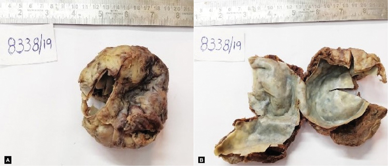
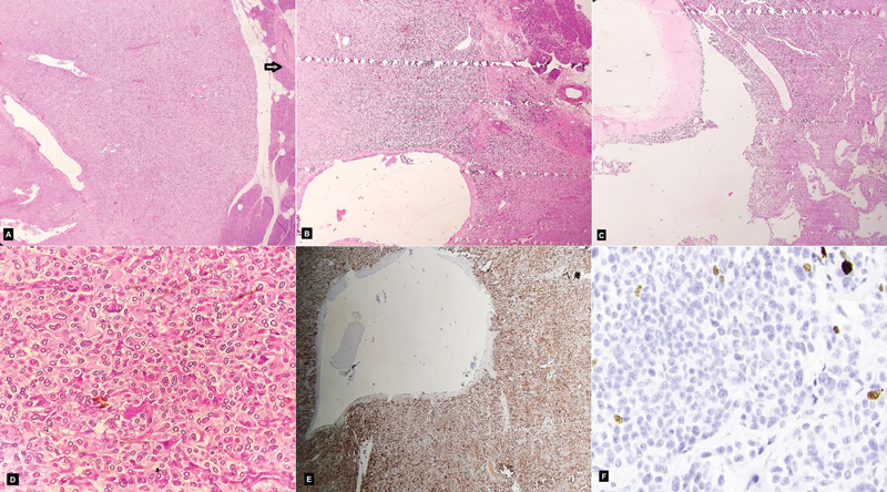
Pancreatic neuroendocrine tumors are typically solid neoplasms but in rare instances may present as cystic lesions. Preoperative diagnosis of a cystic pancreatic lesion is challenging and requires a multidisciplinary and multimodal approach. We hereby describe an elderly female who came with complaints of abdominal lump. Radiologically, it appeared to be a pancreatic hydatid cyst located at the head of the pancreas, following which resection was done. Histopathological study of the lesion turned out to be a cystic pancreatic neuroendocrine tumor. Thus, we present this unique case due to its rarity and diagnostic challenge.
Keywords: chromogranin; cystic lesion; neuroendocrine tumor; pancreas; synaptophysin.
The Indian Association of Laboratory Physicians. This is an open access article published by Thieme under the terms of the Creative Commons Attribution-NonDerivative-NonCommercial License, permitting copying and reproduction so long as the original work is given appropriate credit. Contents may not be used for commercial purposes, or adapted, remixed, transformed or built upon. ( https://creativecommons.org/licenses/by-nc-nd/4.0/ ).
Conflict of interest statement
Conflict of Interest None declared.
Figures
References
-
- Buetow P C, Parrino T V, Buck J L. Islet cell tumors of the pancreas: pathologic-imaging correlation among size, necrosis and cysts, calcification, malignant behavior, and functional status. AJR Am J Roentgenol. 1995;165(05):1175–1179. - PubMed
-
- Kamisawa T, Fukayama M, Koike M, Tabata I, Okamoto A. A case of malignant cystic endocrine tumor of the pancreas. Am J Gastroenterol. 1987;82(01):86–89. - PubMed
-
- Iacono C, Serio G, Fugazzola C. Cystic islet cell tumors of the pancreas. A clinico-pathological report of two nonfunctioning cases and review of the literature. Int J Pancreatol. 1992;11(03):199–208. - PubMed
Publication types
LinkOut - more resources
Full Text Sources