

Urinoma Due to Spontaneous Rupture of the Renal Pelvis Mimicking Appendicitis
- PMID: 37065314
- PMCID: PMC10101196
- DOI: 10.7759/cureus.36141
Urinoma Due to Spontaneous Rupture of the Renal Pelvis Mimicking Appendicitis
Abstract
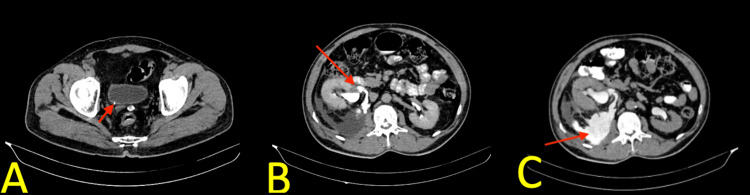
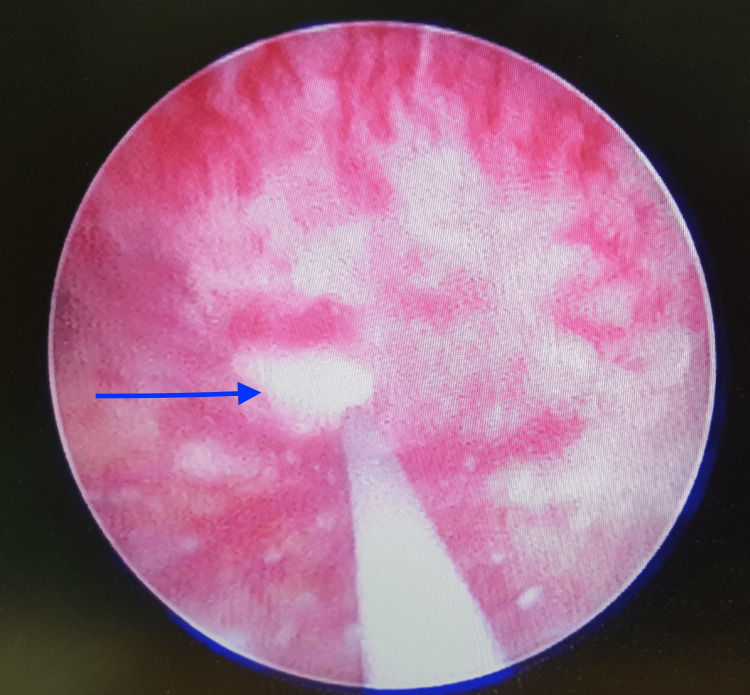
Spontaneous rupture of the renal pelvis (SRRP) with urine extravasation is rare. This condition is primarily associated with an obstructing ureteric calculus. It creates a diagnostic dilemma, especially when the clinical diagnosis can be inconsistent. Herein, we report a 49-year-old male patient who presented with abdominal pain for the past three days and was diagnosed with acute appendicitis. A computed tomography (CT) scan revealed a right renal pelvis rupture and urinoma secondary to an obstructive 4 mm ureterovesical junction calculi. The patient was successfully treated with double-J stent placement. In conclusion, even though SRRP is rare, emergency physicians should have knowledge regarding this condition, which often presents as an abdominal condition and may be misdiagnosed as another condition requiring surgical intervention. Radiologic investigations such as CT scans are useful methods in suspected cases of this condition in order to reduce unnecessary surgical intervention.
Keywords: appendicitis; extravasation; renal pelvic rupture; srrp; urinoma.
Copyright © 2023, Mahawar et al.
Conflict of interest statement
The authors have declared that no competing interests exist.
Figures
Similar articles
-
Spontaneous rupture of renal pelvis as a rare complication of ureteral lithiasis.Turk J Urol. 2016 Mar;42(1):37-40. doi: 10.5152/tud.2015.92979. Turk J Urol. 2016. PMID: 27011880 Free PMC article.
-
Idiopathic Spontaneous Rupture of Renal Pelvis in a Single Functioning Kidney.Case Rep Nephrol Dial. 2021 Jul 22;11(2):221-226. doi: 10.1159/000512588. eCollection 2021 May-Aug. Case Rep Nephrol Dial. 2021. PMID: 34414214 Free PMC article.
-
Spontaneous rupture of the renal pelvis due to obstruction of pelviureteric junction by renal stone: A case report and review of the literature.Urol Ann. 2017 Jul-Sep;9(3):293-295. doi: 10.4103/UA.UA_24_17. Urol Ann. 2017. PMID: 28794602 Free PMC article.
-
Perinephric urinoma following spontaneous renal rupture in the third trimester of pregnancy: a case report and brief review of the literature.BMC Pregnancy Childbirth. 2019 Dec 18;19(1):505. doi: 10.1186/s12884-019-2669-9. BMC Pregnancy Childbirth. 2019. PMID: 31852454 Free PMC article. Review.
-
Spontaneous rupture of the renal pelvis caused by upper urinary tract obstruction: A case report and review of the literature.Medicine (Baltimore). 2017 Dec;96(50):e9190. doi: 10.1097/MD.0000000000009190. Medicine (Baltimore). 2017. PMID: 29390332 Free PMC article. Review.
References
Publication types
LinkOut - more resources
Full Text Sources
Miscellaneous