The Myrtiformis Muscle: Identification of a Forgotten Entity That Is Distinct From the Depressor Septi Nasi Muscle
- PMID: 37065385
- PMCID: PMC10103830
- DOI: 10.7759/cureus.36214
The Myrtiformis Muscle: Identification of a Forgotten Entity That Is Distinct From the Depressor Septi Nasi Muscle
Abstract
Introduction: Nasal musculature anatomy is a topic that plastic surgeons pay attention to. However, the presence and role of the myrtiformis muscle (MM) remain controversial. To elucidate these aspects, an anatomy-based study was conducted.
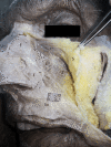
Materials and methods: Seven midsagittally split and two total cadaver head's nasal bases, embalmed with modified Larssen solution (MLS), were dissected for MM anatomy. The features of this muscle were photographed, and a video of its function was recorded.
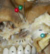
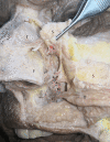
Results: It was found that MM originates from the maxillary alveolar process and continues as two heads, one reaching the alar base with spicular fibrotendinous endings and the other extending to depressor septi nasi fibers. Owing to its bi-vectoral muscle fibers, MM is found to constrict the nares by simultaneously forcing the alar base and lowering the columella. It was also found that left-sided muscles were larger than right-sided muscles.
Conclusions: The MM is found to be a constrictor muscle of the nares in this study, contrary to recent observations.
Keywords: anatomy; cadaver; facial muscle; nose; rhinoplasty.
Copyright © 2023, Yeği̇n et al.
Conflict of interest statement
The authors have declared that no competing interests exist.
Figures


Similar articles
-
Correction of the Topographic Relationship between the Depressor Septi Nasi and Incisivus Labii Superioris: Application to Cosmetic Surgery on the Lip and Nose.Plast Reconstr Surg. 2020 Mar;145(3):524e-529e. doi: 10.1097/PRS.0000000000006558. Plast Reconstr Surg. 2020. PMID: 32097304
-
Revisiting the anatomy of myrtiformis muscle.Surg Radiol Anat. 2023 Jul;45(7):789-794. doi: 10.1007/s00276-023-03154-3. Epub 2023 Apr 27. Surg Radiol Anat. 2023. PMID: 37106241
-
Intranasal approach for manipulating the depressor septi nasi.J Craniofac Surg. 2012 Mar;23(2):367-9. doi: 10.1097/SCS.0b013e318240ca4c. J Craniofac Surg. 2012. PMID: 22421827
-
Multifactorial approaches for correction of the drooping tip of a long nose in East asians.Arch Plast Surg. 2014 Nov;41(6):630-7. doi: 10.5999/aps.2014.41.6.630. Epub 2014 Nov 3. Arch Plast Surg. 2014. PMID: 25396173 Free PMC article. Review.
-
Reconsideration of the alar base cinch suture technique involving the perinasal musculature: an in-depth review.Int J Oral Maxillofac Surg. 2025 Mar;54(3):251-260. doi: 10.1016/j.ijom.2024.07.019. Epub 2024 Aug 16. Int J Oral Maxillofac Surg. 2025. PMID: 39153888 Review.
References
-
- Rhinoplasty. Rohrich RJ, Ahmad J. Plast Reconstr Surg. 2011;128:49–73. - PubMed
-
- Evaluation of methods to reduce formaldehyde levels of cadavers in the dissection laboratory. Whitehead MC, Savoia MC. Clin Anat. 2008;21:75–81. - PubMed
-
- Cadaver embalming fluid for surgical training courses: modified Larssen solution. Bilge O, Celik S. Surg Radiol Anat. 2017;39:1263–1272. - PubMed
-
- The lower nasal base: an anatomical study. Daniel RK, Glasz T, Molnar G, Palhazi P, Saban Y, Journel B. Aesthet Surg J. 2013;33:222–232. - PubMed
-
- Nose muscular dynamics: the tip trigonum. Figallo EE, Acosta JA. Plast Reconstr Surg. 2001;108:1118–1126. - PubMed
LinkOut - more resources
Full Text Sources
