

Comparative Study of Different Entry Spots on Postoperative Gluteus Medius Muscle Cross-Sectional Area in Patients With Intertrochanteric Fractures Nailing
- PMID: 37065393
- PMCID: PMC10097852
- DOI: 10.7759/cureus.36103
Comparative Study of Different Entry Spots on Postoperative Gluteus Medius Muscle Cross-Sectional Area in Patients With Intertrochanteric Fractures Nailing
Abstract
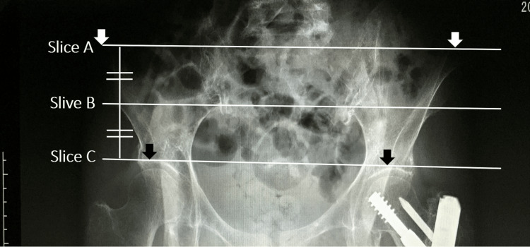
Introduction In a preliminary study of cephalo-medullary (CM) nailing in patients with femoral intertrochanteric fractures, the authors of this study found a 25% to 30% decrease in muscle strength, especially abduction force, during the postoperative follow-up period. This decline was partially attributed to the entry point for the nail insertion causing damage to the gluteus medius tendon at the junction of the greater trochanter after reaming. Therefore, we assumed that changing the position of nail insertion to a "bald spot (BS)" could mitigate postoperative functional impairment. Automated computed tomography (CT) imaging of skeletal muscle cross-sectional area (CSA) and adipose tissue ratio (ATR) can show pathological changes on the operated side compared with the non-operated side. In this study, the authors quantified the difference in postoperative CSA and ATR of the gluteus medius muscle after bald spot nailing versus nail insertion through the conventional tip of the greater trochanter. It was hypothesized that bald spot nailing could avoid significant injury to the gluteus medius muscle. Materials and methods Patients with femoral intertrochanteric fractures were grouped according to the site of cephalo-medullary nailing: greater trochanteric tip (TIP) in 27 patients (8 men and 19 women, mean age 84.9±5.1 years) and BS in 16 patients (3 men and 13 women, mean age 86.9±6.2 years). The CSA and ATR of the gluteus medius muscles were assessed in three slices (A, B, and C from proximal to distal). Each slice was manually traced and automatically calculated based on its contour. Adipose tissue (-100 to -50 in Hounsfield units) in the designated area was distinguished by a bimodal image histogram resulting from the distribution of CT numbers of adipose tissue and muscle. The body mass index (BMI) was used to correct the CSA in each patient. Results In the TIP group, the mean CSA values (mm2) from the non-operated/operated sides were as follows: slice A, 2180.2 ± 616.5/1976.3 ± 421.2; slice B, 2112.3 ± 535.7/1857.7 ± 386.7; and slice C: 1671.8 ± 460.0/1404.1 ± 404.3 (p<0.01 in slices A, B, and C). In the BS group, slice A was 2044.1 ± 473.0/2016.9 ± 388.4; slice B was 2073.2 ± 540.7/1848.3 ± 411.1; and slice C was 1659.1 ± 477.2/1468.5 ± 341.7 (p=0.34 in slice A, and p<0.05 in slices B and C, respectively). The mean CSA values (mm2) of the non-operated minus operated side between the TIP/BS groups were as follows: slice A, 241.3 ± 424.3/-11.8 ± 285.6; slice B, 290.3 ± 313.0/211.8 ± 333.2; and slice C, 276.4 ± 270.4/162.8 ± 319.3 (p < 0.05 in slice A, 0.45, 0.24 in slices B, C, respectively). The mean adjusted CSA per BMI values (mm2) of the non-operated minus the operated side between the TIP/BS groups were slice A, 10.6 ± 19.7/-0.4 ± 14.8; slice B, 13.3 ± 15.0/10.1 ± 16.3; and slice C, 13.1 ± 13.4/ 8.7 ± 15.3 (p < 0.05 in slice A and 0.54 and 0.36 in slices B and C, respectively). Conclusion Nail insertion at the bald spot resulted in a significantly smaller decrease in the CSA of the gluteus medius muscle compared with the conventional tip entry. In addition, an examination of BMI-adjusted CSA showed that CSA was maintained in some image slices. These results suggest that nailing from the BS of the greater trochanter can reduce damage to the gluteus medius muscle and highlight the importance of imaging beyond the usual assessment of skeletal changes.
Keywords: bald spot; cephalo-medullary nailing; comparative study; cross sectional area; entry spot; gamma nailing; gluteus medius; intertrochanteric femur fracture.
Copyright © 2023, Noda et al.
Conflict of interest statement
The authors have declared that no competing interests exist.
Figures


Similar articles
-
Postoperative Gait Performance Following Pertrochanteric Fractures Is Influenced by the Preoperative Condition of the Gluteal Muscles.Cureus. 2024 Aug 30;16(8):e68176. doi: 10.7759/cureus.68176. eCollection 2024 Aug. Cureus. 2024. PMID: 39347225 Free PMC article.
-
Decreased postoperative gluteus medius muscle cross-sectional area measured by computed tomography scan in patients with intertrochanteric fractures nailing.J Orthop Surg (Hong Kong). 2017 Sep-Dec;25(3):2309499017727943. doi: 10.1177/2309499017727943. J Orthop Surg (Hong Kong). 2017. PMID: 28920547
-
Iatrogenic gluteus medius muscle insertion injury while trochanteric entry nailing due to trochanteric fractures: a comparative study in forty patients with gray-scale ultrasound and shear-wave elastography.Int Orthop. 2021 Dec;45(12):3253-3261. doi: 10.1007/s00264-021-05177-0. Epub 2021 Aug 27. Int Orthop. 2021. PMID: 34448922
-
[Nailing of inter- and subtrochanteric fractures - operative technique].Rozhl Chir. 2013 Oct;92(10):615-20. Rozhl Chir. 2013. PMID: 24295486 Review. Czech.
-
Endoscopic Treatment of Gluteus Medius Tears: A Review.Bull Hosp Jt Dis (2013). 2016 Mar;74(1):58-62. Bull Hosp Jt Dis (2013). 2016. PMID: 26977550 Review.
Cited by
-
CT Hounsfield units in assessing bone and soft tissue quality in the proximal femur: A systematic review focusing on osteonecrosis and total hip arthroplasty.PLoS One. 2025 Mar 26;20(3):e0319907. doi: 10.1371/journal.pone.0319907. eCollection 2025. PLoS One. 2025. PMID: 40138288 Free PMC article.
-
The Sonographic Evaluation of Abductor Injury After Intramedullary Nailing for the Hip Fractures.J Clin Med. 2025 Aug 5;14(15):5498. doi: 10.3390/jcm14155498. J Clin Med. 2025. PMID: 40807123 Free PMC article.
-
Postoperative Gait Performance Following Pertrochanteric Fractures Is Influenced by the Preoperative Condition of the Gluteal Muscles.Cureus. 2024 Aug 30;16(8):e68176. doi: 10.7759/cureus.68176. eCollection 2024 Aug. Cureus. 2024. PMID: 39347225 Free PMC article.
References
-
- Gluteus medius tendon injury during reaming for gamma nail insertion. McConnell T, Tornetta P 3rd, Benson E, Manuel J. Clin Orthop Relat Res. 2003:199–202. - PubMed
LinkOut - more resources
Full Text Sources
Miscellaneous