Case Reports
. 2023 Mar 13;15(3):e36098.
doi: 10.7759/cureus.36098. eCollection 2023 Mar.

A Case of Septic Pulmonary Embolism Caused by Pyelonephritis With Klebsiella pneumoniae in a Patient With Poorly Controlled Type 2 Diabetes Mellitus

Affiliations
Case Reports

A Case of Septic Pulmonary Embolism Caused by Pyelonephritis With Klebsiella pneumoniae in a Patient With Poorly Controlled Type 2 Diabetes Mellitus

Miho Katsumata et al. Cureus. .

Abstract

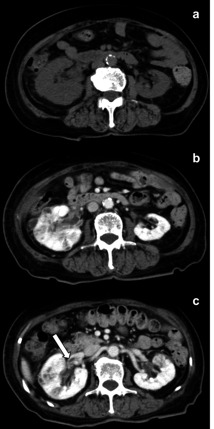
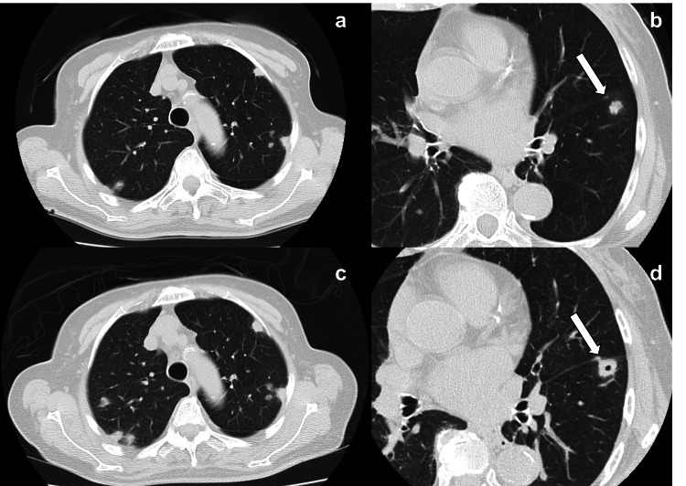
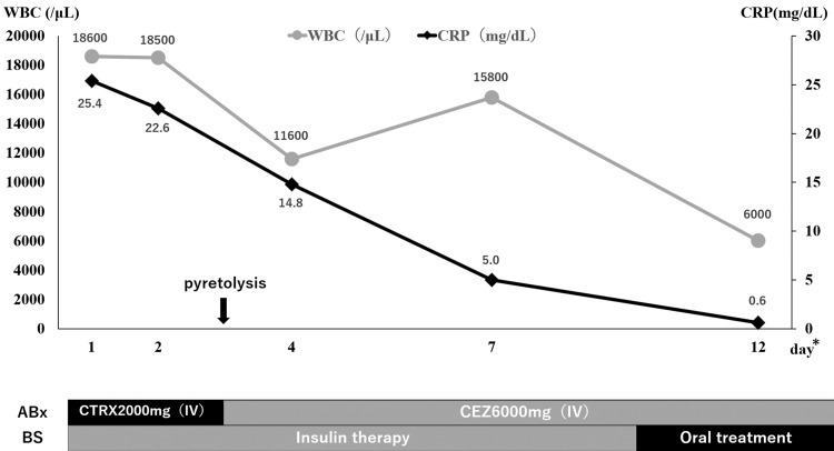
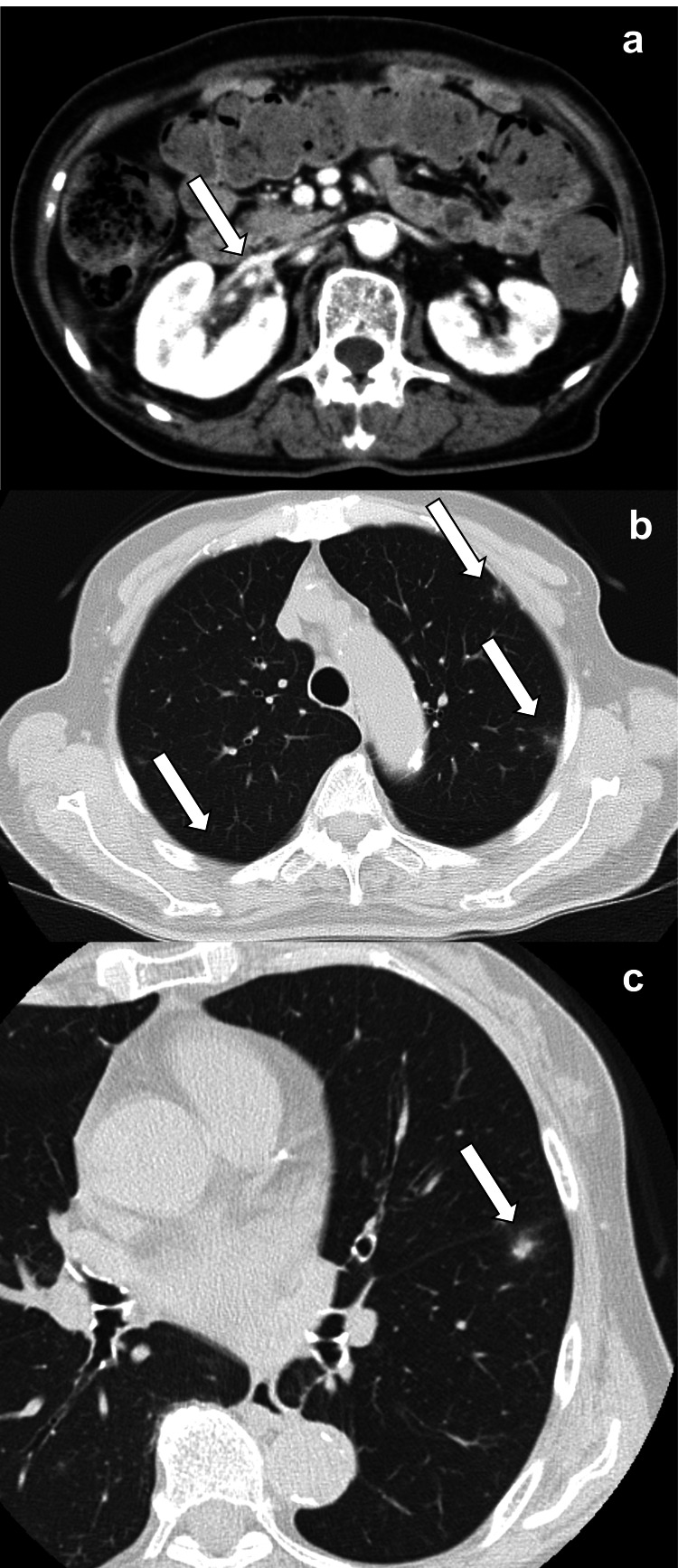
Septic pulmonary embolism (SPE) is caused by the microbe that is responsible for any clinical condition that may include urinary tract infections as in this case. We report a case of pyelonephritis with Klebsiella pneumoniae that led to SPE in an 80-year-old woman with poorly controlled diabetes mellitus (DM). Computed tomography (CT) revealed multiple nodules in the peripheral area of the bilateral lung and a contrast defect in the right renal vein, which was suspected to be an embolism. Blood and urine cultures revealed Klebsiella pneumoniae infection. These results confirmed the diagnosis of pyelonephritis and SPE. Treatment with ceftriaxone, cefazolin, and ciprofloxacin improved the patient's condition.

Keywords: klebsiella pneumoniae (kp); poorly controlled diabetes; septic pulmonary emboli; type 2 diabetes mellitus (dm); urinary tract infection.


Conflict of interest statement

The authors have declared that no competing interests exist.

Figures

Figure 1
Figure 1. Image of chest radiograph on admission
Multiple nodules in the upper bilateral lobe. The arrows indicate pulmonary nodules.
Figure 2
Figure 2. Images of abdominal CT
Bilateral renal swelling and perinephric standing are also observed (a). Poor contrast enhancements in bilateral renal parenchyma are observed (b). Suspected bacterial mass in the right vein. The arrow indicates a suspected bacterial mass (c).
Figure 3
Figure 3. Images of chest CT
Multiple nodules are observed in the peripheral area on admission (a, b). Multiple nodules are enlarged and cavities appeared in some of them on day 4 (c, d). The arrows indicate a nodule with enlargement and a cavity.
Figure 4
Figure 4. Process of treatment and inflammatory markers
The symbol * indicates days of hospitalization. The treatment was initiated with ceftriaxone 2 g per day. After confirming bacterial species and antimicrobial susceptibility, it was de-escalated to cefazolin 6 g per day IV on day 3. Insulin was also initiated to control blood glucose and then switched to oral medication. After treatment initiation, the patient's temperature resolved, and inflammatory markers rapidly decreased. WBC, white blood cell; CRP, C-reactive protein; ABx, antibiotics; BS, blood sugar; CTRX, ceftriaxone; CEZ, cefazolin; IV, intravenous.
Figure 5
Figure 5. One-month follow-up CT after treatment initiation
Bilateral renal swelling improved, and poor contrast enhancement regressed, and the arrow indicates the point at which a suspected bacterial mass was detected on day 4 (a). Multiple nodules and cavities regressed or disappeared. The arrows indicate nodules and cavities with regression and disappearance (b, c).

Similar articles

References

    1. Septic pulmonary embolism: presenting features and clinical course of 14 patients. Cook RJ, Ashton RW, Aughenbaugh GL, Ryu JH. Chest. 2005;128:162–166. - PubMed
    1. The association of septic thrombophlebitis with septic pulmonary embolism in adults. Brenes JA, Goswami U, Williams DN. Open Respir Med J. 2012;6:14–19. - PMC - PubMed
    1. Klebsiella pneumoniae infection associated septic pulmonary embolism in an emergency department from east China. Zhang X, Yang Q, Gao B, et al. Ann Palliat Med. 2021;10:1521–1529. - PubMed
    1. Pulmonary septic emboli: diagnosis with CT. Kuhlman JE, Fishman EK, Teigen C. Radiology. 1990;174:211–213. - PubMed
    1. Computed tomography findings in septic pulmonary embolism: a case report and literature review. Owji S, Choi WJ, Al-Jabbari E, Manral K, Palacio D, Bhargava P. Radiol Case Rep. 2022;17:2639–2642. - PMC - PubMed

Publication types

LinkOut - more resources