

Primary hepatic paraganglioma with megacolon: A case report
- PMID: 37065786
- PMCID: PMC10091477
- DOI: 10.3892/ol.2023.13769
Primary hepatic paraganglioma with megacolon: A case report
Abstract
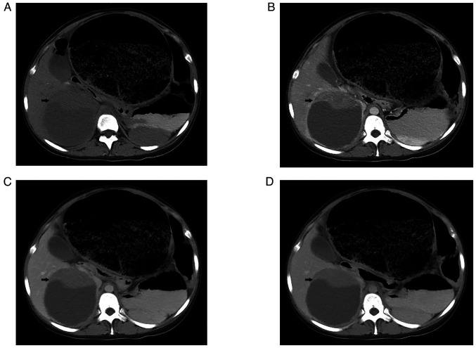
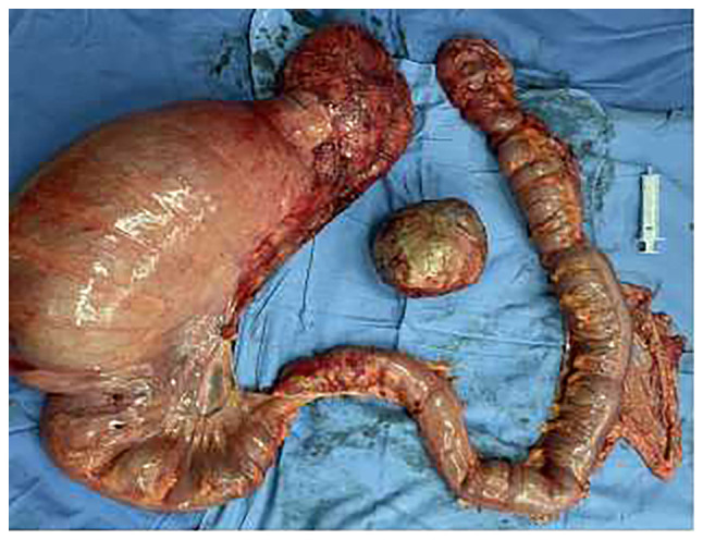
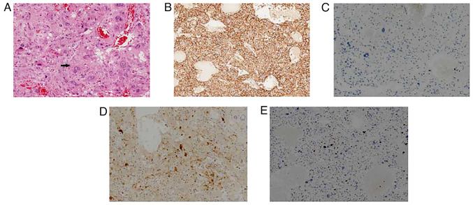
Primary hepatic paraganglioma (PGL) is a rare neuroendocrine tumor characterized by clinical manifestations including paroxysmal hypertension, palpitation, abdominal pain and constipation. In the present study, the case of a 21-year-old woman with pathologically confirmed hepatic PGL with megacolon following surgery is reported. The patient initially visited Beijing Tiantan Hospital (Beijing, China) for hypoferric anemia. A triple-phase CT scan of the whole abdomen showed a large hypodense mass with a solid periphery and strong arterial enhancement of the peripheral solid portion of the liver. The sigmoid colon and rectum were obviously distended, filled with gas and intestinal contents. The patient was preoperatively diagnosed with iron deficiency anemia, liver injury and megacolon and then underwent partial hepatectomy, total colectomy and enterostomy. Microscopically, the liver cells exhibited an irregular zellballen pattern. In addition, immunohistochemical staining revealed that liver cells were positive for CD56, chromogranin A, vimentin, S-100, melan-A and neuron-specific enolase. Therefore, the diagnosis of primary PGL of the liver was confirmed. These findings suggested that primary hepatic PGL should not be excluded when megacolon occurs and comprehensive imaging evaluation is of great importance for its diagnosis.
Keywords: catecholamine; hepatic paraganglioma; megacolon; zellballen.
Copyright: © Bo et al.
Conflict of interest statement
The authors declare that they have no competing interests.
Figures
Similar articles
-
A primary paraganglioma of the liver mimicking hepatocellular carcinoma.Tzu Chi Med J. 2019 Sep 16;31(4):286-288. doi: 10.4103/tcmj.tcmj_230_18. eCollection 2019 Oct-Dec. Tzu Chi Med J. 2019. PMID: 31867261 Free PMC article.
-
Primary hepatic paraganglioma mimicking hepatocellular carcinoma: a case report.Transl Cancer Res. 2022 Sep;11(9):3434-3439. doi: 10.21037/tcr-22-314. Transl Cancer Res. 2022. PMID: 36237251 Free PMC article.
-
Paraganglioma of the urinary bladder initially diagnosed as gastrointestinal stromal tumor requiring combined resection of the rectum: a case report.World J Surg Oncol. 2022 Jun 8;20(1):185. doi: 10.1186/s12957-022-02662-7. World J Surg Oncol. 2022. PMID: 35676716 Free PMC article.
-
Primary pancreatic paraganglioma: a case report and literature review.World J Surg Oncol. 2016 Jan 22;14(1):19. doi: 10.1186/s12957-016-0771-2. World J Surg Oncol. 2016. PMID: 26801079 Free PMC article. Review.
-
A rare case of multiple paragangliomas in the head and neck, retroperitoneum and duodenum: A case report and review of the literature.Front Endocrinol (Lausanne). 2023 Jan 10;13:1054468. doi: 10.3389/fendo.2022.1054468. eCollection 2022. Front Endocrinol (Lausanne). 2023. PMID: 36704041 Free PMC article. Review.
Cited by
-
Perioperative Management Challenges in Silent Pheochromocytoma: A Case Report and Literature Review.Clin Case Rep. 2025 Apr 6;13(4):e70396. doi: 10.1002/ccr3.70396. eCollection 2025 Apr. Clin Case Rep. 2025. PMID: 40196048 Free PMC article.
References
-
- Lloyd RV, Osamura YR, Kloppel G, Rosa J, editors. WHO Classification of Tumours. 4th edition. Vol. 10. World Health Organization; Geneva: 2017. WHO classification of tumours of endocrine organs.
-
- Corti B, D'Errico A, Pierangeli F, Fiorentino M, Altimari A, Grigioni WF. Primary paraganglioma strictly confined to the liver and mimicking hepatocellular carcinoma: An immunohistochemical and in situ hybridization study. Am J Surg Pathol. 2002;26:945–949. doi: 10.1097/00000478-200207000-00015. - DOI - PubMed
-
- Khan MR, Raza R, Jabbar A, Ahmed A. Primary non-functioning paraganglioma of liver: A rare tumour at an unusual location. J Pak Med Assoc. 2011;6:814–816. - PubMed
Publication types
LinkOut - more resources
Full Text Sources
Research Materials