. 2023 Mar 31:11:1104620.
doi: 10.3389/fcell.2023.1104620. eCollection 2023.

Probing the conserved roles of cut in the development and function of optically different insect compound eyes

Affiliations

Probing the conserved roles of cut in the development and function of optically different insect compound eyes

Shubham Rathore et al. Front Cell Dev Biol. .

Abstract

Astonishing functional diversity exists among arthropod eyes, yet eye development relies on deeply conserved genes. This phenomenon is best understood for early events, whereas fewer investigations have focused on the influence of later transcriptional regulators on diverse eye organizations and the contribution of critical support cells, such as Semper cells (SCs). As SCs in Drosophila melanogaster secrete the lens and function as glia, they are critical components of ommatidia. Here, we perform RNAi-based knockdowns of the transcription factor cut (CUX in vertebrates), a marker of SCs, the function of which has remained untested in these cell types. To probe for the conserved roles of cut, we investigate two optically different compound eyes: the apposition optics of D. melanogaster and the superposition optics of the diving beetle Thermonectus marmoratus. In both cases, we find that multiple aspects of ocular formation are disrupted, including lens facet organization and optics as well as photoreceptor morphogenesis. Together, our findings support the possibility of a generalized role for SCs in arthropod ommatidial form and function and introduces Cut as a central player in mediating this role.

Keywords: compound eyes; cone cells; conserved gene networks; invertebrates; optics; semper cells; visual system development.


Conflict of interest statement

The authors declare that the research was conducted in the absence of any commercial or financial relationships that could be construed as a potential conflict of interest.

Figures

FIGURE 1
FIGURE 1
Compound eye types and development. (A) D. melanogaster has a neural-superposition eye, the optics of which follows typical apposition organization, with individual lenses (L) that each project a tiny image fragment onto the tips of underlying photoreceptor rhabdomeres (R). Underneath the lens, there is a pseudocone (PC) and four Semper cell (SC) bodies. (B) T. marmoratus has an optical superposition eye, in which sets of lenses synergistically project image points onto corresponding underlying closed rhabdoms. In this organization, the SC bodies are located in close proximity to the lens and above the photoreceptor cell bodies. The optics require the presence of pronounced crystalline cones (CCs) and a clear zone. (C) Compound eye development is best understood in D. melanogaster, in which specific cell types are sequentially recruited from a precursor epithelium. Top: Diagram of cell fate specification and differentiation in the Drosophila compound eye. Bottom: The four SCs within each ommatidium show Cut immunoreactivity (green) in the larval, pupal, and adult stages. For better orientation, counterstained tissue is illustrated in magenta: ELAV in larvae, E-cadherin in pupae, and drosocrystallin (lenses) in adults.
FIGURE 2
FIGURE 2
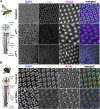
Cut expression and RNAi-driven knockdown in D. melanogaster and T. marmoratus compound eyes. (A). In D. melanogaster, a quartet of Cut-positive Semper cells (green) are situated within the distal-most portion of developing ommatidia (at ∼37% pupal development). DAPI and N-cadherin counterstaining are used to identify the correct layer within the eye. As illustrated in representative images, efficient cut knockdown is achieved by two different SC-directed RNAis, with overlapping phenotypes consisting of an irregular ommatidial array. Incidences of laterally displaced rhabdoms are indicated via N-cadherin staining (arrowhead). (B). In T. marmoratus, at a comparable developmental stage, four Cut-positive SCs are similarly organized near the distal margin of each ommatidium. At this stage, the closed rhabdom (red; confirmed by actin staining) still resides in close proximity to the SCs. The nuclear localization of Cut is confirmed by complete overlap with DAPI. cutRNAi treated individuals show a strong but incomplete reduction of Cut, with irregularities in the ommatidial array. In some instances at this level, only a triad of Cut-positive nuclei are visible (arrow), and rhabdoms appear to be laterally displaced (arrowhead). Scale bars = 10 µm.
FIGURE 3
FIGURE 3
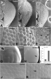
Cut knockdown affects lens organization in insect compound eyes. (A–F) Scanning electron micrographs of adult D. melanogaster compound eyes. Overview of a control individual (A) illustrates a typical completely regular ommatidium array, whereas ct GD (B) and ct V20 (C) exhibit major irregularities in ommatidial placement and lens formation. The latter is illustrated in a magnified view of the anterior region of the compound eye. In control individuals (D), lenses appear precisely shaped with properly formed lens surfaces. In ct GD (E) and ct V20 (F), irregularities exist in ommatidium separation, with some neighboring units fused (arrows). In some instances, the lens surface exhibits deformities typical of the blueberry phenotype (arrowheads), which are particularly pronounced in the ct V20 line. (G–K) Scanning electron micrographs of adult T. marmoratus compound eyes. Overview of a control beetle (G) shows an intact eye with a smooth surface. In cutRNAi individuals (H), surface dimples (arrow) are more common than in controls (I). A high-resolution image of the anterior region of the compound eye illustrates precise placement and smooth transitions between neighboring ommatidia (J), whereas cutRNAi injected individuals show irregularities in ommatidium size and more delineated borders (K). Additionally, some neighboring units are fused (arrow). Scale bars = 100 µm (A–C), 40 µm (D–F), 500 µm (G, H), and 50 µm (J, K).
FIGURE 4
FIGURE 4
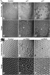
Morphological lens defects lead to profound optical deficits. Isolated lens arrays were used to visualize the back surfaces of lenses (A–F) and images of an object with three stripes were then produced by these lens arrays (A′–F′). In D. melanogaster, control lenses have smooth and accurately formed back surfaces (A) that lead to regularly spaced and equally sized images with a uniform focal plane across the lens array (A′). In contrast, the lenses of the cut knockdown lines show visible defects in morphology (B, C) and optics, with images that vary in placement, image magnification (arrowheads), focal plane, and blurriness (B′, C′). For ct V20, several lenses show dimple-like indentations on the back surfaces (arrowhead) and other lenses appear dark and necrotic (C). Such necrotic lenses (exemplified by the cluster marked with *) lead to gaps in the resulting image array * in (C′). In T. marmoratus, a similar pattern is observed, with controls having smooth and even lens back surfaces (D) that result in pristine regular image arrays (D′). In contrast, cutRNAi individuals exhibit lens irregularities (E) that lead to irregularities in the corresponding lens array (E′), including greatly displaced images (arrowhead). Lens back surfaces frequently show dimple-like lens indentations arrowheads in (E, F), which are also present in individuals with fewer irregularities in lens placement (F). Even in this morphologically less severe phenotype, major deficiencies in the lens array optics occur, resulting in many blurry images and some differently sized images (arrowhead) (F′). Scale bars = 50 µm.
FIGURE 5
FIGURE 5
Cut knockdown leads to rhabdom misplacement in both eye types. (A). In D. melanogaster, rhabdoms typically extend along the majority of the ommatidia, from close to the pseudocone to the basement membrane. (B). Rhabdoms, visualized with phalloidin, appear well developed and regular in control individuals. In ct GD (C) and ct V20 (D) individuals, rhabdoms appear truncated and frequently misplaced, with many extending well below the basement membrane (arrowheads). (E). In T. marmoratus, rhabdoms are situated much deeper in the eye to make room for a clear zone, which is necessary to allow many lenses to contribute to the image formed at the distal end of the PRs. (F). Phalloidin staining in control individuals illustrates precisely aligned rhabdoms that extend from below the clear zone to well above the basement membrane (BM). PR nuclei are aligned precisely along a concentric circle between the rhabdoms and lenses. (G). In cutRNAi individuals, PR placement is less regular, at the levels of both PR nuclei and rhabdoms. As in D. melanogaster, rhabdoms are displaced toward the basement membrane and occasionally traverse it (arrowhead). Scale bars = 50 µm (B–D) and 100 µm (F, G).
FIGURE 6
FIGURE 6
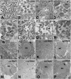
Cut knockdown leads to ultrastructural defects of rhabdoms in both eye types. (A). As illustrated by a control individual, D. melanogaster has an open rhabdom that, at any cross-sectional plane, is formed by seven rhabdomeres. (B). At higher magnification, it is apparent that the smaller central rhabdomere extends into the center of an extracellular lumen, which is bordered by larger and approximately evenly sized outer rhabdomeres. (C). Overview of a ct GD knockdown individual illustrates ommatidial displacements (with a compromised interommatidial space) and deformed or missing rhabdomeres (exemplified by the unit marked with *). (D). Several units characterized by relatively extended or even split rhabdomeres (arrowhead). (E). Other units showing unusually small rhabdomeres (arrowhead). (F). Overview of a ct V20 knockdown individual illustrates ommatidia with relatively sparse rhabdomeres, large extracellular spaces between rhabdomeres, and sparse and degenerate interommatidial tissue. Non-etheless, ct V20 individuals also show laterally extended rhabdoms (G), arrow, split rhabdomeres (G), arrowhead, and possibly fused rhabdomeres (H), arrow. (I). As illustrated by a control individual, the superposition eyes of T. marmoratus are characterized by closed rhabdoms (two units with similar rhabdom diameters in close proximity are marked with *). (J) The rhabdom is positioned centrally within a healthy ommatidium. (K). In cutRNAi individuals, neighboring units (marked with *) show relatively different rhabdom organization. (L) An unusually shaped rhabdom with central deficiencies. (M) A laterally displaced and strongly degenerate rhabdom. (N) Overview of several ommatidia in a different individual shows the complete absence of a rhabdom (^) next to two neighboring semi-intact rhabdoms (*). (O) A laterally degenerate rhabdom. (P) A rhabdom with a displaced portion (arrowhead). Scale bars = 5 µm (A,C, F), 2 µm (B, D, E, G, H, J, L, M, O, P), and 10 µm (I, K, N); Rh = rhabdomere (B, D) or rhabdom (J, M, O, P).
FIGURE 7
FIGURE 7
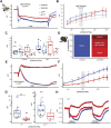
Despite major structural deficits, electroretinograms of cut knockdown individuals show relatively intact physiological responses in D. melanogaster and relatively minor deficiencies in T. marmoratus. (A). Example recordings from two control and two test flies illustrate comparable responses. (B). Average responses (with standard error) to increasing light intensities suggest a comparable dynamic range across the four tested fly lines (n = 10 each). (C). Example responses at two different light intensities. (D). Quantification of cutRNAi injected beetles shows inverted responses at all (dark red) or higher (medium red) light intensities. (E). Example recordings of a control individual and one of each of the three phenotypes in (D). (F). Average responses (with standard error) to increasing light intensities suggest a comparable dynamic range between control and cutRNAi individuals, albeit with generally lower responses in the knockdowns (*p < 0.05, **p < 0.005; based on Wilcoxon’s rank sum test). (G). Example responses at two different light intensities. (H). Example of cutRNAi individuals showing different response dynamics when multiple pulses are presented.
FIGURE 8
FIGURE 8
Schematic summary of SC-mediated effects of cut.

Similar articles

Cited by

References

    1. Agi E., Langen M., Altschuler S. J., Wu L. F., Zimmermann T., Hiesinger P. R. (2014). The evolution and development of neural superposition. J. Neurogenetics 28, 216–232. 10.3109/01677063.2014.922557 - DOI - PMC - PubMed
    1. Bauke A-C., Sasse S., Matzat T., Klämbt C. (2015). A transcriptional network controlling glial development in the Drosophila visual system. Development 142, 2184–2193. 10.1242/dev.119750 - DOI - PubMed
    1. Belušič G. (2011). “ERG in Drosophila,” in Electroretinograms. Editor Belušič G. (London: IntecOpen; ). 10.5772/21747 - DOI
    1. Blackie L., Walther R. F., Staddon M. F., Banerjee S., Pichaud F. (2020). Cell-type-specific mechanical response and myosin dynamics during retinal lens development in Drosophila . Mol. Biol. Cell. 31, 1355–1369. 10.1091/mbc.E19-09-0523 - DOI - PMC - PubMed
    1. Blackie L., Tozluoglu M., Trylinski M., Walther R. F., Schweisguth F., Mao Y., et al. (2021). A combination of Notch signaling, preferential adhesion and endocytosis induces a slow mode of cell intercalation in the Drosophila retina. Development 148, 197301. 10.1242/dev.197301 - DOI - PMC - PubMed

LinkOut - more resources