

Leiomyoma of the fallopian tube found during laparoscopic myomectomy: A case report and review of the literature
- PMID: 37066002
- PMCID: PMC10102355
- DOI: 10.3389/fsurg.2023.997338
Leiomyoma of the fallopian tube found during laparoscopic myomectomy: A case report and review of the literature
Abstract
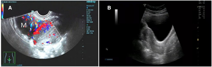
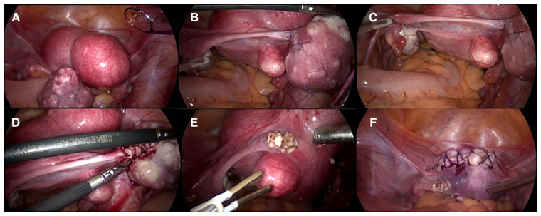
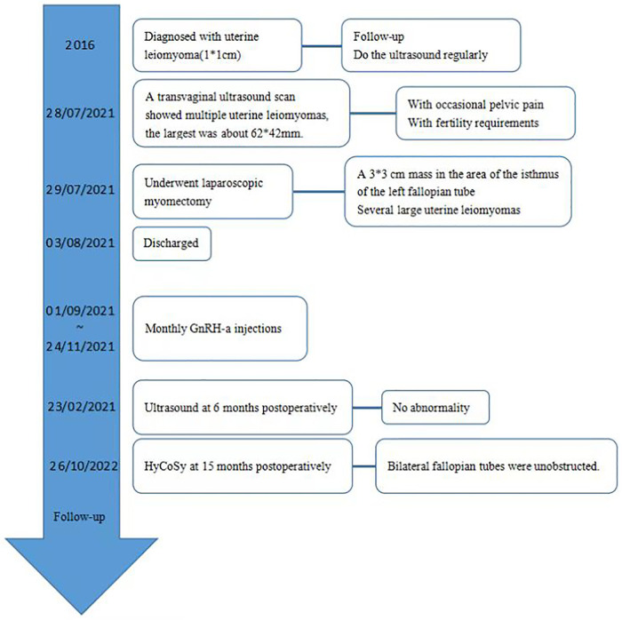
Leiomyoma of the fallopian tube is an extremely rare benign tumor of the fallopian tube. Because of the small number of cases, it is difficult to calculate their incidence. In this case report, we report a case of leiomyoma of the fallopian tube detected during laparoscopic myomectomy in a 31-year-old female with occasional pelvic pain. The patient was diagnosed with uterine leiomyoma based on a transvaginal ultrasound scan. She was operated and a 3*3 cm mass in the area of the isthmus of the left fallopian tube was observed. Three uterine leiomyomas and one leiomyoma of the fallopian tube were removed. Ultrasound at 6 months postoperatively showed no abnormality. Hysterosalpingo-contrast-sonography (HyCoSy) at 15 months postoperatively showed bilateral fallopian tubes were unobstructed. For those patients with fertility requirements, some fertility-preserving techniques can be used to allow complete resection of the leiomyoma and avoid tubal damage.
Keywords: benign tumors; fallopian tube neoplasms; fertility; laparoscopic myomectomy; leiomyoma.
© 2023 Cheng, Wang, Fu and Fu.
Conflict of interest statement
The authors declare that the research was conducted in the absence of any commercial or financial relationships that could be construed as a potential conflict of interest.
Figures
Similar articles
-
[TORSION OF FALLOPIAN TUBE LEIOMYOMA TREATED BY LAPAROSCOPY].Akush Ginekol (Sofiia). 2016;55(2):49-51. Akush Ginekol (Sofiia). 2016. PMID: 27509659 Bulgarian.
-
Leiomyoma and leiomyoma cellulare of the fallopian tube: review of the literature and case reports.Prz Menopauzalny. 2016 Nov;15(3):143-147. doi: 10.5114/pm.2016.63053. Epub 2016 Nov 15. Prz Menopauzalny. 2016. PMID: 27980525 Free PMC article.
-
Combined Real-Time Three-Dimensional Hysterosalpingo-Contrast Sonography with B Mode Hysterosalpingo-Contrast Sonography in the Evaluation of Fallopian Tube Patency in Patients Undergoing Infertility Investigations.Biomed Res Int. 2019 Jun 3;2019:9408141. doi: 10.1155/2019/9408141. eCollection 2019. Biomed Res Int. 2019. PMID: 31275995 Free PMC article.
-
Primary leiomyoma of the fallopian tube: preoperative ultrasound findings.J Chin Med Assoc. 2007 Feb;70(2):80-3. doi: 10.1016/S1726-4901(09)70307-7. J Chin Med Assoc. 2007. PMID: 17339150 Review.
-
Port site parasitic leiomyoma after laparoscopic myomectomy: a case report and review of the literature.J Med Case Rep. 2018 Nov 15;12(1):339. doi: 10.1186/s13256-018-1873-y. J Med Case Rep. 2018. PMID: 30428912 Free PMC article. Review.
Cited by
-
A Massive Adenoma of the Uterine Tube in a Young Intact Female Dog: Surgical Intervention and Outcome.Vet Sci. 2025 Mar 7;12(3):253. doi: 10.3390/vetsci12030253. Vet Sci. 2025. PMID: 40266977 Free PMC article.
References
Publication types
LinkOut - more resources
Full Text Sources