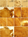
Synaptic connectivity of the TRPV1-positive trigeminal afferents in the rat lateral parabrachial nucleus
- PMID: 37066077
- PMCID: PMC10098450
- DOI: 10.3389/fncel.2023.1162874
Synaptic connectivity of the TRPV1-positive trigeminal afferents in the rat lateral parabrachial nucleus
Abstract
Recent studies have shown a direct projection of nociceptive trigeminal afferents into the lateral parabrachial nucleus (LPBN). Information about the synaptic connectivity of these afferents may help understand how orofacial nociception is processed in the LPBN, which is known to be involved primarily in the affective aspect of pain. To address this issue, we investigated the synapses of the transient receptor potential vanilloid 1-positive (TRPV1+) trigeminal afferent terminals in the LPBN by immunostaining and serial section electron microscopy. TRPV1 + afferents arising from the ascending trigeminal tract issued axons and terminals (boutons) in the LPBN. TRPV1+ boutons formed synapses of asymmetric type with dendritic shafts and spines. Almost all (98.3%) TRPV1+ boutons formed synapses with one (82.6%) or two postsynaptic dendrites, suggesting that, at a single bouton level, the orofacial nociceptive information is predominantly transmitted to a single postsynaptic neuron with a small degree of synaptic divergence. A small fraction (14.9%) of the TRPV1+ boutons formed synapses with dendritic spines. None of the TRPV1+ boutons were involved in axoaxonic synapses. Conversely, in the trigeminal caudal nucleus (Vc), TRPV1+ boutons often formed synapses with multiple postsynaptic dendrites and were involved in axoaxonic synapses. Number of dendritic spine and total number of postsynaptic dendrites per TRPV1+ bouton were significantly fewer in the LPBN than Vc. Thus, the synaptic connectivity of the TRPV1+ boutons in the LPBN differed significantly from that in the Vc, suggesting that the TRPV1-mediated orofacial nociception is relayed to the LPBN in a distinctively different manner than in the Vc.
Keywords: lateral parabrachial nucleus; nociceptive; synaptic connectivity; trigeminal; ultrastructure.
Copyright © 2023 An, Cho, Park, Kim and Bae.
Conflict of interest statement
The authors declare that the research was conducted in the absence of any commercial or financial relationships that could be construed as a potential conflict of interest. The handling editor S-YC declared a past co-authorship with the author YB.
Figures


Similar articles
-
Ultrastructural analysis of the synaptic connectivity of TRPV1-expressing primary afferent terminals in the rat trigeminal caudal nucleus.J Comp Neurol. 2010 Oct 15;518(20):4134-46. doi: 10.1002/cne.22369. J Comp Neurol. 2010. PMID: 20878780
-
Synaptic connectivity of urinary bladder afferents in the rat superficial dorsal horn and spinal parasympathetic nucleus.J Comp Neurol. 2019 Dec 15;527(18):3002-3013. doi: 10.1002/cne.24725. Epub 2019 Jun 12. J Comp Neurol. 2019. PMID: 31168784
-
Expression of transient receptor potential ankyrin 1 (TRPA1) in the rat trigeminal sensory afferents and spinal dorsal horn.J Comp Neurol. 2010 Mar 1;518(5):687-98. doi: 10.1002/cne.22238. J Comp Neurol. 2010. PMID: 20034057
-
GABA distribution in a pain-modulating zone of trigeminal subnucleus interpolaris.Somatosens Res. 1988;5(3):205-17. doi: 10.3109/07367228809144627. Somatosens Res. 1988. PMID: 2895952 Review.
-
Ultrastructural basis for craniofacial sensory processing in the brainstem.Int Rev Neurobiol. 2011;97:99-141. doi: 10.1016/B978-0-12-385198-7.00005-9. Int Rev Neurobiol. 2011. PMID: 21708309 Review.
Cited by
-
Structural reorganization of medullary dorsal horn astrocytes in a rat model of neuropathic pain.Brain Struct Funct. 2024 Sep;229(7):1757-1768. doi: 10.1007/s00429-024-02835-y. Epub 2024 Jul 25. Brain Struct Funct. 2024. PMID: 39052094
-
Sensorimotor regulation of facial expression - An untouched frontier.Neurosci Biobehav Rev. 2024 Jul;162:105684. doi: 10.1016/j.neubiorev.2024.105684. Epub 2024 May 6. Neurosci Biobehav Rev. 2024. PMID: 38710425 Free PMC article. Review.
-
Role of TRP Channels in Cancer-Induced Bone Pain.Int J Mol Sci. 2025 Jan 30;26(3):1229. doi: 10.3390/ijms26031229. Int J Mol Sci. 2025. PMID: 39940997 Free PMC article. Review.
-
Effect of chronic intermittent hypoxia on ocular and intraoral mechanical allodynia mediated via the calcitonin gene-related peptide in a rat.Sleep. 2024 Mar 11;47(3):zsad332. doi: 10.1093/sleep/zsad332. Sleep. 2024. PMID: 38166171 Free PMC article.
References
-
- Bae Y. C., Ihn H. J., Park M. J., Ottersen O. P., Moritani M., Yoshida A., et al. (2000). Identification of signal substances in synapses made between primary afferents and their associated axon terminals in the rat trigeminal sensory nuclei. J. Comp. Neurol. 418 299–309. - PubMed
LinkOut - more resources
Full Text Sources

