[Preprint]. 2023 Apr 7:2023.04.06.535968.
doi: 10.1101/2023.04.06.535968.

Interferon ε restricts Zika virus infection in the female reproductive tract

Affiliations

Interferon ε restricts Zika virus infection in the female reproductive tract

Chuan Xu et al. bioRxiv. .

Update in

  • Interferon ɛ restricts Zika virus infection in the female reproductive tract.
    Xu C, Wang A, Ebraham L, Sullivan L, Tasker C, Pizutelli V, Couret J, Hernandez C, Kolli P, Deb PQ, Fritzky L, Subbian S, Gao N, Lo Y, Salvatore M, Rivera A, Lemenze A, Fitzgerald-Bocarsly P, Tyagi S, Lu W, Beaulieu A, Chang TL. Xu C, et al. PNAS Nexus. 2023 Oct 25;2(11):pgad350. doi: 10.1093/pnasnexus/pgad350. eCollection 2023 Nov. PNAS Nexus. 2023. PMID: 37954158 Free PMC article.

Abstract

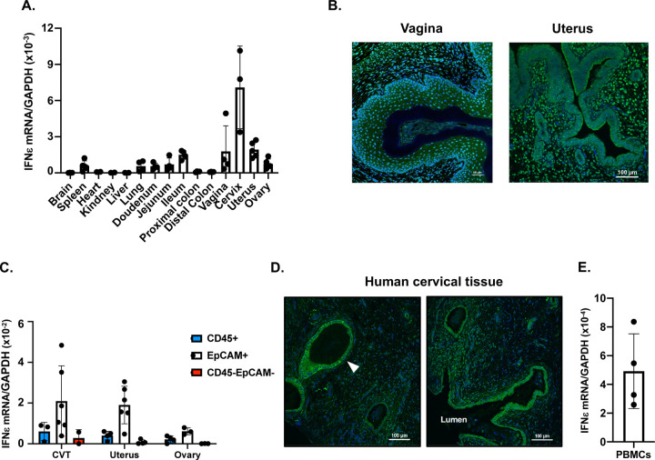
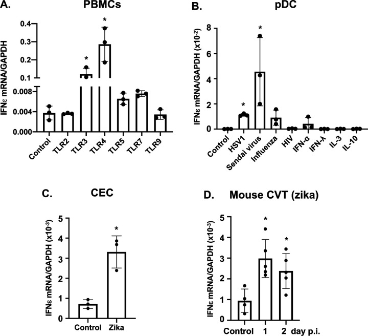
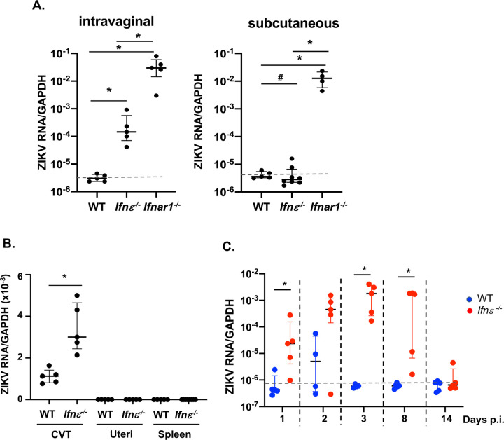
Interferon ε (IFNε) is a unique type I IFN that has been implicated in host defense against sexually transmitted infections (STIs). Zika virus (ZIKV), an emerging pathogen, can infect the female reproductive tract (FRT) and cause devastating diseases, particularly in pregnant women. How IFNε contributes to protection against ZIKV infection in vivo is unknown. Here, we show that IFNε plays a critical role in host protection against vaginal ZIKV infection in mice. We found that IFNε was expressed not only by epithelial cells in the FRT, but also by certain immune and other cells at baseline or after exposure to viruses or specific TLR agonists. IFNε-deficient mice exhibited abnormalities in the epithelial border and underlying tissue in the cervicovaginal tract, and these defects were associated with increased susceptibility to vaginal, but not subcutaneous ZIKV infection. IFNε-deficiency resulted in an increase in magnitude, duration, and depth of ZIKV infection in the FRT. Critically, intravaginal administration of recombinant IFNε protected Ifnε-/- mice and highly susceptible Ifnar1-/- mice against vaginal ZIKV infection, indicating that IFNε was sufficient to provide protection even in the absence of signals from other type I IFNs and in an IFNAR1-independent manner. Our findings reveal a potentially critical role for IFNε in mediating protection against transmission of ZIKV in the context of sexual contact.


Conflict of interest statement

The authors have declared that no conflict of interest exists.

Figures

Figure 1.
Figure 1.. IFNε is expressed in epithelial and non-epithelial cells at the female reproductive tract.
(A) Total RNAs were extracted from different tissues of wild-type (WT) mice. IFNε mRNA levels were quantified by RT-qPCR and normalized by GAPDH using the 2−ΔΔCT method. Each point represents one animal. (B) Expression of IFNε protein (Green) in vaginal (left) and uterus (right) tissue. Nuclei were stained with DAPI (blue). (C) IFNε mRNA levels were quantified by RT-qPCR of total RNA from sorted CD45+ cells, epithelial cells (EpCAM), and CD45EpCAM cells from cervicovaginal tissue (CVT), uterus, and ovary. (D) Detection of human IFNε proteins in human cervical tissues by immunofluorescence staining. The arrow indicates glandular epithelial cells expressing IFNε proteins. (E) Total RNAs were extracted from freshly isolated PBMCs from different donors. IFNε mRNA levels were quantified by RT-qPCR.
Figure 2.
Figure 2.. IFNε is induced in response to viral infection and TLR activation.
(A) Freshly isolated PBMCs from different donors were stimulated with agonists for TLR2 (Pam3CSK4,100 ng/ml), TLR3 (poly (I:C), 1 μg/ml), TLR4 (LPS, 10 ng/ml), TLR5 (FLA-ST, 50 ng/ml), TLR7 (Imiquimod, 500 ng/ml), or TLR9 (ODN2006, 5 μM) for 6 h. Total RNAs were prepared and IFNε gene expression was quantified by RT-qPCR. (B) Plasmacytoid dendritic cells (pDCs) from different donors were purified using a negative selection pDC isolation kit. Purified pDCs were untreated (control) or treated with HSV1 2931, Sendai VR 907, influenza virus PR/8/34, HIV MN, rIFNα, IFNλ, IL-3, or IL-10 for 4 h. Total RNAs were prepared, and IFNε mRNA levels were quantified by RT-qPCR. (C) IFNε was determined by RT-qPCR in primary cervical epithelial cells (CEC) 24 h after ZIKV infection. (D) Induction of IFNε gene expression in the cervicovaginal tract of WT mice in response to ZIKV infection. *p<0.5, treated vs untreated controls.
Figure 3.
Figure 3.. Ifnε−/− mice exhibit aberrant epithelial and tissue structure in the cervicovaginal tissue (CVT).
(A) Ifnε−/− mice were generated using CRISPR/Cas9 technology. Knockdown of IFNε gene expression in CVT, uteri (U), and ovaries (O) of Ifnε−/− mice was confirmed by RT-qPCR. (B) IFNε protein expression in WT and Ifnε−/− mice was determined by IHC. IFNε proteins (brown) were found in the vaginal tissue of WT mice but not of Ifnε−/− mice. (C, D) WT and Ifnε−/− mice were synchronized to the diestrus stage by Depo-Provera. Total RNAs from the CVT were collected on day 12 after injection. RNAseq analyses showing pathways (C) and genes (D) involved in epithelial integrity, tissue structure, and wound healing in CVT of Ifnε−/− mice. (E) A schematic diagram of murine FRT. The site of sectioning for collagens and epithelial structure examination are indicated. (F) Collagens in the cervix of WT and Ifnε−/− mice were stained with Picro Sirius Red. Cervical canal (C) and vaginal fornix (VF) are indicated. (G) Higher power views (20x) of vaginal epithelial folds and surrounding collagen structure. Black arrows show the difference in collagen organization. (H) The length of vaginal epithelial folds in mid regions of vaginal epithelial fornix in WT and Ifnε−/− mice.
Figure 4.
Figure 4.. Ifnε−/− mice exhibit increased susceptibility to intravaginal ZIKV infection.
(A) Depo-Provera-synchronized WT, Ifnε−/−, and Ifnar1−/− mice were challenged with ZIKV PRVABC59 through an intravaginal or subcutaneous route. Zika RNA levels at the CVT were determined by RT-qPCR at day 3 p.i. Data represent 5 independent experiments. (B) Depo-Provera-treated WT and Ifnε−/− mice were infected by zika virus intravaginally. Total RNAs from CVT, uteri, and spleen were harvested at day 3 p.i. Zika RNA levels were determined by RT-qPCR. Data represent 3 experiments (C) Depo-Provera-treated WT and Ifnε−/− mice were challenged with ZIKV intravaginally. Total RNAs of the CVT were prepared at different days p.i. ZIKV signals were determined by RT-qPCR. Data represent 3 experiments. The dash line indicates the background from uninfected mice. *p<0.05; ns, not significant.
Figure 5.
Figure 5.. Faster dissemination of ZIKV of Ifnε−/− mice
(A) A serial sections of the CVT from uterus fundus toward the distal cervix. H&E staining of the proximal and mid cervix of WT and of Ifnε−/− mice used for smFISH are shown. (B). Adjacent sections to the H&E-stained sections were used for smFISH to detect ZIKV RNAs (red). Images at 20x are shown. Nuclei are in blue and autofluorescence is in green. Identical exposures were used for all images including both WT and Ifnε−/− mice. (C). Localization of ZIKA signals in Ifnε−/− mice (62x) in lamina propria (left) and stroma (right).
Figure 6.
Figure 6.. Intravaginal administration of murine IFNε proteins protect mice against ZIKV infection.
(A) Depo-Provera synchronized Ifnε−/− mice were treated with recombinant mIFNε proteins (4 μg) through an intravaginal route for 6 h followed by ZIKV infection. ZIKV RNA levels at CVT were determined by RT-qPCR at day 3 p.i. The dash line indicates the background from uninfected control. (B) Depo-Provera synchronized Ifnar1−/− mice were treated with recombinant mIFNε proteins (4 μg) through an intravaginal or subcutaneous route for 6 h followed by zika infection via the same route. Zika RNA levels at the CVT and spleen were determined by RT-qPCR at day 3 p.i. (C) Depo-Provera-synchronized Ifnar1−/− mice were treated with PBS or linearized mIFNε proteins (4 μg) for 6 h intravaginally followed by intravaginal ZIKV infection. Viral RNA levels at the CVT were detected by RT-qPCR. (D) Depo-Provera-synchronized Ifnar1−/− mice were treated with mIFNε proteins (4 μg) intravaginally 6 h before or one day after (D1) intravaginal ZIKV infection. Viral RNA levels at the CVT were detected by RT-qPCR. *p<0.05, #p>0.05. The values of the background from uninfected Ifnar1−/− mice were 1–3×10⁻⁶. Data represent 3–4 independent experiments.

Similar articles

References

    1. Pavlidis I. et al., Cervical epithelial damage promotes Ureaplasma parvum ascending infection, intrauterine inflammation and preterm birth induction in mice. Nat Commun 11, 199 (2020). - PMC - PubMed
    1. Lacroix G., Gouyer V., Gottrand F., Desseyn J. L., The Cervicovaginal Mucus Barrier. Int J Mol Sci 21 (2020). - PMC - PubMed
    1. Lozenski K., Ownbey R., Wigdahl B., Kish-Catalone T., Krebs F. C., Decreased cervical epithelial sensitivity to nonoxynol-9 (N-9) after four daily applications in a murine model of topical vaginal microbicide safety. BMC Pharmacol Toxicol 13, 9 (2012). - PMC - PubMed
    1. Catalone B. J. et al., Mouse model of cervicovaginal toxicity and inflammation for preclinical evaluation of topical vaginal microbicides. Antimicrob Agents Chemother 48, 1837–1847 (2004). - PMC - PubMed
    1. Fichorova R. N., Desai P. J., Gibson F. C. 3rd, Genco C. A., Distinct proinflammatory host responses to Neisseria gonorrhoeae infection in immortalized human cervical and vaginal epithelial cells. Infect Immun 69, 5840–5848 (2001). - PMC - PubMed

Publication types