[Preprint]. 2023 Apr 6:2023.04.06.535927.
doi: 10.1101/2023.04.06.535927.

SARS-CoV-2 Mac1 is required for IFN antagonism and efficient virus replication in mice

Affiliations

SARS-CoV-2 Mac1 is required for IFN antagonism and efficient virus replication in mice

Yousef M Alhammad et al. bioRxiv. .

Update in

Abstract

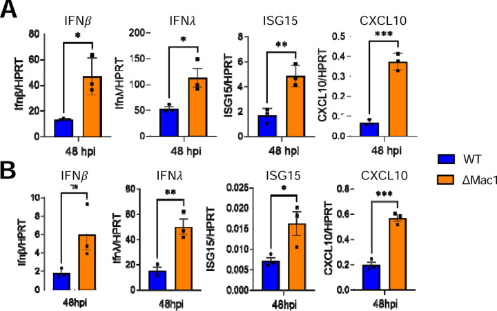
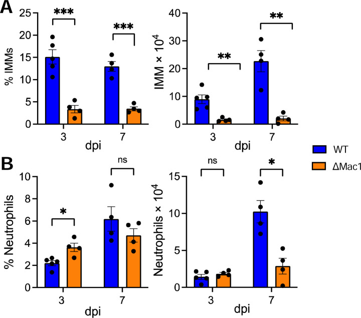
Several coronavirus (CoV) encoded proteins are being evaluated as targets for antiviral therapies for COVID-19. Included in this set of proteins is the conserved macrodomain, or Mac1, an ADP-ribosylhydrolase and ADP-ribose binding protein. Utilizing point mutant recombinant viruses, Mac1 was shown to be critical for both murine hepatitis virus (MHV) and severe acute respiratory syndrome (SARS)-CoV virulence. However, as a potential drug target, it is imperative to understand how a complete Mac1 deletion impacts the replication and pathogenesis of different CoVs. To this end, we created recombinant bacterial artificial chromosomes (BACs) containing complete Mac1 deletions (ΔMac1) in MHV, MERS-CoV, and SARS-CoV-2. While we were unable to recover infectious virus from MHV or MERS-CoV ΔMac1 BACs, SARS-CoV-2 ΔMac1 was readily recovered from BAC transfection, indicating a stark difference in the requirement for Mac1 between different CoVs. Furthermore, SARS-CoV-2 ΔMac1 replicated at or near wild-type levels in multiple cell lines susceptible to infection. However, in a mouse model of severe infection, ΔMac1 was quickly cleared causing minimal pathology without any morbidity. ΔMac1 SARS-CoV-2 induced increased levels of interferon (IFN) and interferon-stimulated gene (ISG) expression in cell culture and mice, indicating that Mac1 blocks IFN responses which may contribute to its attenuation. ΔMac1 infection also led to a stark reduction in inflammatory monocytes and neutrophils. These results demonstrate that Mac1 only minimally impacts SARS-CoV-2 replication, unlike MHV and MERS-CoV, but is required for SARS-CoV-2 pathogenesis and is a unique antiviral drug target.

Significance: All CoVs, including SARS-CoV-2, encode for a conserved macrodomain (Mac1) that counters host ADP-ribosylation. Prior studies with SARS-CoV-1 and MHV found that Mac1 blocks IFN production and promotes CoV pathogenesis, which has prompted the development of SARS-CoV-2 Mac1 inhibitors. However, development of these compounds into antivirals requires that we understand how SARS-CoV-2 lacking Mac1 replicates and causes disease in vitro and in vivo . Here we found that SARS-CoV-2 containing a complete Mac1 deletion replicates normally in cell culture but induces an elevated IFN response, has reduced viral loads in vivo , and does not cause significant disease in mice. These results will provide a roadmap for testing Mac1 inhibitors, help identify Mac1 functions, and open additional avenues for coronavirus therapies.


Conflict of interest statement

Competing interests: The University of Kansas-Lawrence has filed a patent application relating to coronavirus live-attenuated vaccines which lists A.R.F and R.C. as co-inventors.

Figures

Fig. 1.
Fig. 1.. SARS-CoV-2 Mac1 deletion virus replicates normally in Vero E6 and A549-ACE2 cells.
VeroE6 (A-B) and A549-ACE2 (C) cells were infected with SARS-CoV-2 WT and ∆Mac1 at an MOI of 0.1 (A,C) and 1 (B) PFU/cell. Both cell-associated and cell-free virus was collected at indicated time points and virus-titers were determined by plaque assays. Data shown is one experiment representative of three independent experiments. n = 3 per group for each experiment. ns – not significant.
Fig. 2.
Fig. 2.. SARS-CoV-2 has a mild replication defect in Calu-3 cells.
(A-D) Calu-3 cells were infected with SARS-CoV-2 WT and Mac1 viruses at both low (A-B) and high (C-D) MOI. Both cell-associated and cell-free virus was collected at indicated times and virus titers were determined by plaque assay. The data in A & C are from one experiment representative of at least 3 independent experiments. n = 3 per group. The results of all combined experiments where the average WT values from each experiment were normalized to 1.0 at 24 and 48 hpi are shown in B and D. Each point represents a separate biological replicate. (E-F) Calu-3 cells were infected at an MOI of 1 PFU/cell as described above and cell lysates were collected, and viral protein levels were determined by immunoblotting (E) or cells fixed at 24 hpi were co-stained with DAPI and either anti-nsp3 or anti-N, and then analyzed by confocal microscopy at 20X magnification (F). The data in E-F shows data from one representative experiment of two independent experiments.
Fig. 3.
Fig. 3.. IFN-γ, but not IFN-β, pretreatment enhances replication defect of Mac1 in Calu-3 cells.
Calu-3 cells were pretreated for 18 h with increasing concentrations (0, 5, 50, and 500 units) of IFN-β (A) and IFN-γ (B), then infected with either SARS-CoV-2 WT or ∆Mac1 at an MOI of 0.1 PFU/cell. Cells were collected at 48 hpi and titers were determined by plaque assay. Fold differences between WT and Mac1 are indicated at each amount of IFN. The data shown are of one experiment representative of two (A) and three (B) independent experiments. n = 3 for each group.
Fig. 4.
Fig. 4.. Mac1 induces increased IFN and cytokines responses compared to WT SARS-CoV-2 in cell culture.
Calu3 (A) and A549-ACE2 (B) cells were infected with SARS-CoV-2 WT and ∆Mac1 at an MOI of 0.1 PFU/cell and total RNA was collected 48 hpi. IFN-β, IFN-λ, ISG15 and CXCL10 levels were determined by qPCR using ∆Ct method with primers listed in table S2 and normalized to HPRT mRNA levels. The data show one experimental representative of three independent experiments with n = 3 for each experiment.
Fig. 5.
Fig. 5.. Mac1 is highly attenuated in K18-ACE2 mice.
(A-B) K18-ACE2 C57BL/6 mice were infected with 2.5×10⁴ PFU of WT or ∆Mac1 SARS-CoV-2 and survival and weight loss were measured over 12 days. (C) Photomicrographs (hematoxylin and eosin stain) of lungs from WT and ∆Mac1 infected mice at 7 dpi demonstrating bronchointerstitial pneumonia (black arrow) and edema and fibrin (black asterisk) (D) Mice were scored for bronchointerstitial pneumonia, inflammation, and edema/fibrin deposition. WT n=4; ∆Mac1 n=5. (E-F) K18-ACE2 C57BL/6 mice were infected as described above and lung titers (E) and gRNA levels (F) were determined by plaque assay and RT-qPCR with primers specific for nsp12 and normalized to HPRT, respectively. Results are from one experiment representative of two independent experiments. n = 4–10 mice per group.
Fig. 6.
Fig. 6.. Mac1 virus induces a robust innate immune response in the lungs following infection.
(A) K18-ACE2 C57BL/6 mice were infected with 2.5×10⁴ PFU of indicated viruses and lungs were harvested at 1 dpi and total RNA was isolated. The relative levels of indicated transcripts were determined by qPCR using the ∆Ct method with primers listed in table S2 normalized to HPRT mRNA levels. The results are from one experiment representative of two independent experiments with an n = 4–8 mice per group. (B-D) The total RNA from the samples in (A) were analyzed by RNAseq to determine the full transcriptome in the lung following infection. (B) Volcano plot indicating differentially expressed genes (DEGs) between WT and ∆Mac1 infected mice. (C) Functional enrichment analysis of biological processes enriched in the transcriptome of in mice infected with ∆Mac1 performed using DAVID functional annotation tool. (D) Log2 fold change values of genes involved in innate immune response upregulated in mice infected with ∆Mac1 compared to WT virus.
Fig. 7.
Fig. 7.. Mac1 virus infection results in reduced inflammatory monocytes and neutrophils.
(A-B) K18-ACE2 C57BL/6 mice were infected as described above and lungs were harvested at the indicated days post-infection, and the percentages and total numbers of infiltrating inflammatory monocytes (A) and neutrophils (B) were determined by flow cytometry. Data are derived from the results of 1 experiment representative of 2 independent experiments performed with 4–5 mice/group/experiment.

References

    1. Wang Y., Grunewald M., Perlman S., Coronaviruses: An Updated Overview of Their Replication and Pathogenesis. Methods Mol Biol 2203, 1–29 (2020). - PMC - PubMed
    1. Blanco-Melo D. et al., Imbalanced Host Response to SARS-CoV-2 Drives Development of COVID-19. Cell 181, 1036–1045 e1039 (2020). - PMC - PubMed
    1. Parker E. P. K. et al., Response to additional COVID-19 vaccine doses in people who are immunocompromised: a rapid review. Lancet Glob Health 10, e326–e328 (2022). - PMC - PubMed
    1. Beigel J. H., Tomashek K. M., Dodd L. E., Remdesivir for the Treatment of Covid-19 - Preliminary Report. Reply. N Engl J Med 383, 994 (2020). - PubMed
    1. Wahl A. et al., SARS-CoV-2 infection is effectively treated and prevented by EIDD-2801. Nature 591, 451–457 (2021). - PMC - PubMed

Publication types