[Preprint]. 2023 Apr 8:2023.04.07.536026.
doi: 10.1101/2023.04.07.536026.

The Chromatin Landscape of Pathogenic Transcriptional Cell States in Rheumatoid Arthritis

Affiliations

The Chromatin Landscape of Pathogenic Transcriptional Cell States in Rheumatoid Arthritis

Kathryn Weinand et al. bioRxiv. .

Update in

  • The chromatin landscape of pathogenic transcriptional cell states in rheumatoid arthritis.
    Weinand K, Sakaue S, Nathan A, Jonsson AH, Zhang F, Watts GFM, Al Suqri M, Zhu Z; Accelerating Medicines Partnership Program: Rheumatoid Arthritis and Systemic Lupus Erythematosus (AMP RA/SLE) Network; Rao DA, Anolik JH, Brenner MB, Donlin LT, Wei K, Raychaudhuri S. Weinand K, et al. Nat Commun. 2024 May 31;15(1):4650. doi: 10.1038/s41467-024-48620-7. Nat Commun. 2024. PMID: 38821936 Free PMC article.

Abstract

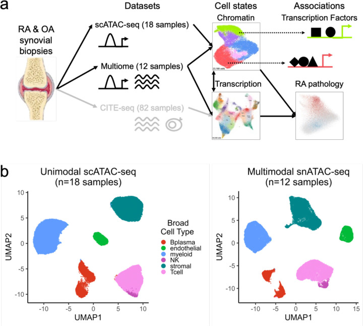
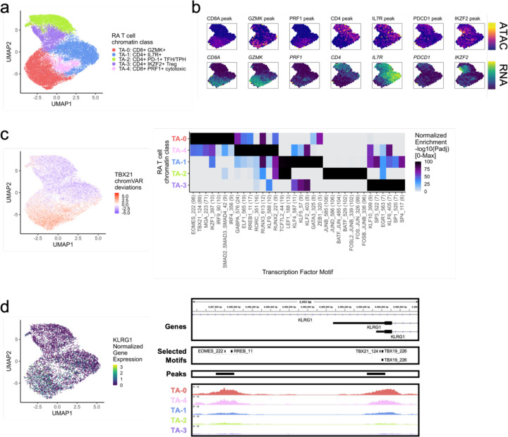
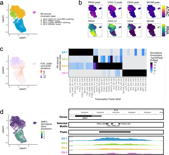
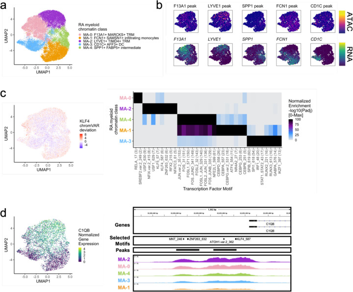
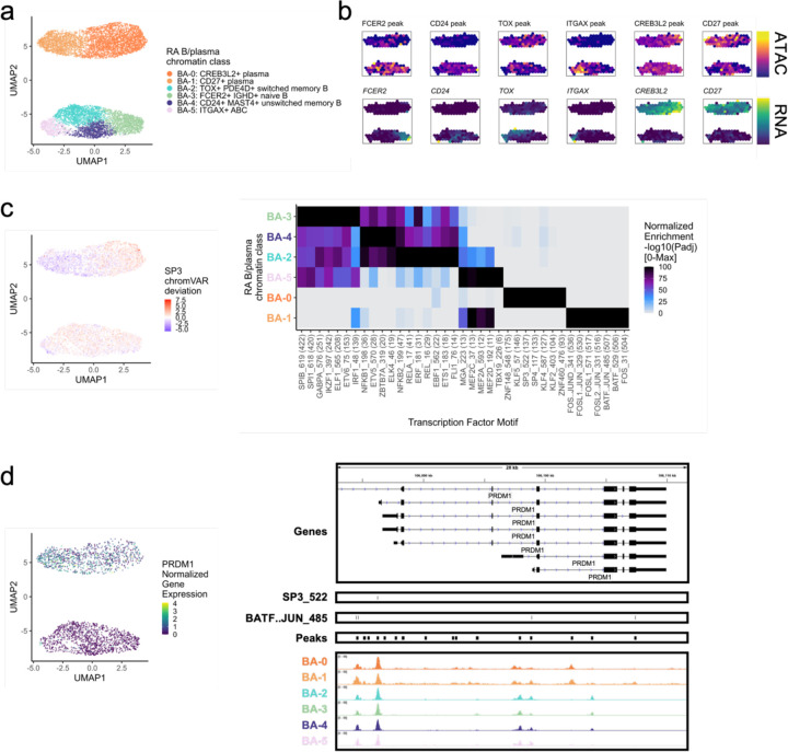
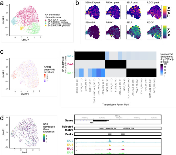
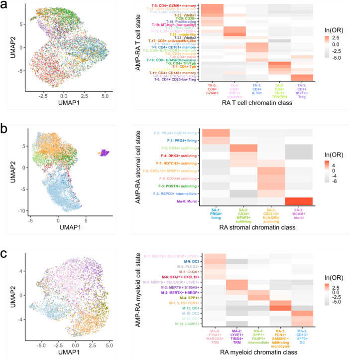
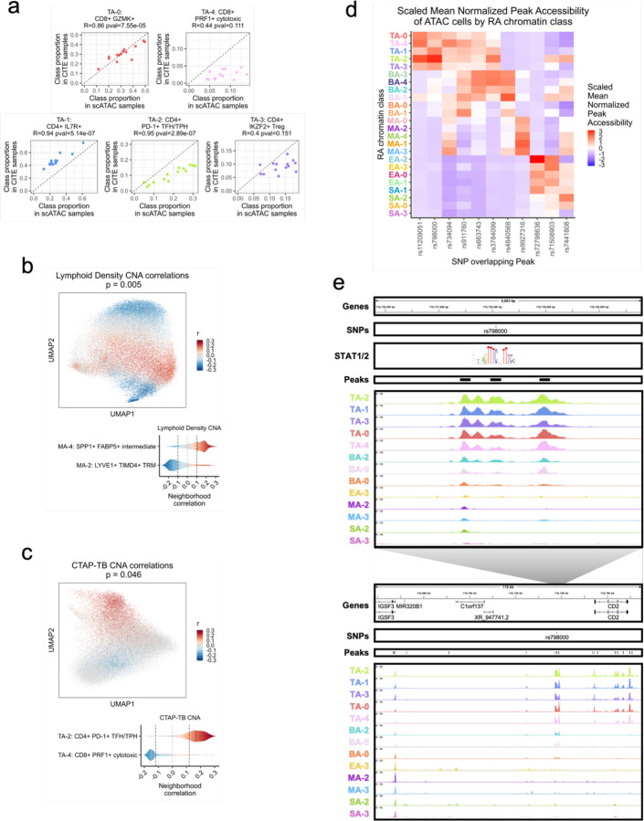
Synovial tissue inflammation is the hallmark of rheumatoid arthritis (RA). Recent work has identified prominent pathogenic cell states in inflamed RA synovial tissue, such as T peripheral helper cells; however, the epigenetic regulation of these states has yet to be defined. We measured genome-wide open chromatin at single cell resolution from 30 synovial tissue samples, including 12 samples with transcriptional data in multimodal experiments. We identified 24 chromatin classes and predicted their associated transcription factors, including a CD8+ GZMK+ class associated with EOMES and a lining fibroblast class associated with AP-1. By integrating an RA tissue transcriptional atlas, we found that the chromatin classes represented 'superstates' corresponding to multiple transcriptional cell states. Finally, we demonstrated the utility of this RA tissue chromatin atlas through the associations between disease phenotypes and chromatin class abundance as well as the nomination of classes mediating the effects of putatively causal RA genetic variants.


Conflict of interest statement

Competing Interests S.R. is a founder for Mestag Therapeutics, a scientific advisor for Janssen and Pfizer, and a consultant for Gilead. D.A.R. reports personal fees from Pfizer, Janssen, Merck, GlaxoSmithKline, AstraZeneca, Scipher Medicine, HiFiBio, and Bristol-Myers Squibb, and grant support from Merck, Janssen, and Bristol-Myers Squibb outside the submitted work. D.A.R. is a co-inventor on the patent for Tph cells as a biomarker of autoimmunity.

Figures

Fig. 1.
Fig. 1.
Study overview and open chromatin broad cell type identification. a. Study overview. Synovial biopsies from RA and OA patients were utilized for unimodal scATAC-seq, multimodal snATAC-seq + snRNA-seq experiments. CITE-seq was performed in the AMP-RA reference study. We defined chromatin classes using the unimodal and multimodal ATAC data and compared them with AMP-RA transcriptional cell states classified onto the multiome cells. We further defined transcription factors likely regulating these chromatin classes and found putative links to RA pathology by associating the classes to RA clinical metrics, RA subtypes, and putative RA risk variants. b. Open chromatin broad cell type identification in unimodal scATAC-seq datasets (left) and multimodal snATAC-seq datasets (right), processed separately.
Fig. 2.
Fig. 2.
RA T cell chromatin classes. a. UMAP colored by 5 T cell chromatin classes defined from unimodal scATAC and multimodal snATAC cells. b. Binned normalized marker peak accessibility (top) and gene expression (bottom) for multiome snATAC cells on UMAP. c. UMAP colored by chromVAR deviations for the TBX21 motif (left). Most significantly enriched motifs in marker peaks per T cell chromatin class (right). To be included per class, motifs had to be enriched in the class above a minimal threshold and corresponding TFs had to have at least minimal expression in snRNA (Methods). Color scale normalized per motif across classes with max −log10(padj) value shown in parentheses in motif label. P-values were calculated via hypergeometric test in ArchR. d. UMAP colored by KLRG1 normalized gene expression in multiome cells (left). KLRG1 locus (chr12:8,987,550-8,990,000) with selected isoforms, motifs, open chromatin peaks, and chromatin accessibility reads from unimodal and multimodal ATAC cells aggregated by chromatin class and scaled by read counts per class (Methods) (right).
Fig. 3.
Fig. 3.
RA stromal chromatin classes. a. UMAP colored by 4 stromal chromatin classes defined from unimodal scATAC and multimodal snATAC cells. b. Binned normalized marker peak accessibility (top) and gene expression (bottom) for multiome snATAC cells on UMAP. c. UMAP colored by chromVAR deviations for the FOS..JUND motif (left). Most significantly enriched motifs in marker peaks per stromal chromatin class (right). To be included per class, motifs had to be enriched in the class above a minimal threshold and corresponding TFs had to have at least minimal expression in snRNA (Methods). Color scale normalized per motif across classes with max −log10(padj) value shown in parentheses in motif label. P-values were calculated via hypergeometric test in ArchR. d. UMAP colored by MMP3 normalized gene expression (left). MMP3 locus (chr11:102,843,400-102,844,000) with selected isoforms, motifs, open chromatin peaks, and chromatin accessibility reads from unimodal and multimodal ATAC cells aggregated by chromatin class and scaled by read counts per class (Methods) (right).
Fig. 4.
Fig. 4.
RA myeloid chromatin classes. a. UMAP colored by 5 myeloid chromatin classes defined from unimodal scATAC and multimodal snATAC cells. b. Binned normalized marker peak accessibility (top) and gene expression (bottom) for multiome snATAC cells on UMAP. c. UMAP colored by chromVAR deviations for the KLF4 motif (left). Most significantly enriched motifs in marker peaks per myeloid chromatin class (right). To be included per class, motifs had to be enriched in the class above a minimal threshold and corresponding TFs had to have at least minimal expression in snRNA (Methods). Color scale normalized per motif across classes with max −log10(padj) value shown in parentheses in motif label. P-values were calculated via hypergeometric test in ArchR. d. UMAP colored by C1QB normalized gene expression (left). C1QB locus (chr1:22,652,235-22,653,595) with selected isoforms, motifs, open chromatin peaks, and chromatin accessibility reads from unimodal and multimodal ATAC cells aggregated by chromatin class and scaled by read counts per class (Methods) (right).
Fig. 5.
Fig. 5.
RA B/plasma chromatin classes. a. UMAP colored by 6 B/plasma chromatin classes defined from unimodal scATAC and multimodal snATAC cells. b. Binned normalized marker peak accessibility (top) and gene expression (bottom) for multiome snATAC cells on UMAP. c. UMAP colored by chromVAR deviations for the SP3 motif (left). Most significantly enriched motifs in marker peaks per B/plasma chromatin class (right). To be included per class, motifs had to be enriched in the class above a minimal threshold and corresponding TFs had to have at least minimal expression in snRNA (Methods). Color scale normalized per motif across classes with max −log10(padj) value shown in parentheses in motif label. P-values were calculated via hypergeometric test in ArchR. d. UMAP colored by PRDM1 normalized gene expression (left). PRDM1 locus (chr6:106,082,865-106,111,658) with selected isoforms, motifs, open chromatin peaks, and chromatin accessibility reads from unimodal and multimodal ATAC cells aggregated by chromatin class and scaled by read counts per class (Methods) (right).
Fig. 6.
Fig. 6.
RA endothelial chromatin classes. a. UMAP colored by 4 endothelial chromatin classes defined from unimodal scATAC and multimodal snATAC cells. b. Binned normalized marker peak accessibility (top) and gene expression (bottom) for multiome snATAC cells on UMAP. c. UMAP colored by chromVAR deviations for the SOX17 motif (left). Most significantly enriched motifs in marker peaks per endothelial chromatin class (right). To be included per class, motifs had to be enriched in the class above a minimal threshold and corresponding TFs had to have at least minimal expression in snRNA (Methods). Color scale normalized per motif across classes with max −log10(padj) value shown in parentheses in motif label. P-values were calculated via hypergeometric test in ArchR. EA-3 is not shown because only 1 marker peak was found, likely due to low cell counts. d. UMAP colored by NES normalized gene expression (left). NES locus (chr1:156,675,399-156,680,400) with selected isoforms, motifs, open chromatin peaks, and chromatin accessibility reads from unimodal and multimodal ATAC cells aggregated by chromatin class and scaled by read counts per class (Methods) (right).
Fig. 7.
Fig. 7.
A chromatin class encompassed multiple transcriptional cell states in proposed superstate model. For (a.) T, (b.) stromal, and (c.) myeloid cells, UMAP colored by classified AMP-RA reference transcriptional cell states for multiome cells (left) and natural log of Odds Ratio between chromatin classes and transcriptional cell states (right). Non-significant values (FDR<0.05) are white. In c., M-13: pDC transcriptional cell state was excluded as fewer than 10 cells were classified into it.
Fig. 8.
Fig. 8.
Linking RA chromatin classes to RA pathology. a. For each donor shared between the unimodal ATAC and AMP-RA reference studies with at least 200 T cells, the Pearson correlation between the relative proportions of T cell chromatin classes defined in the unimodal ATAC datasets (x-axis) and classified into in the CITE datasets through the multiome cells (y-axis). Pearson Correlation Coefficients (R) and p-values (pval) noted. b. CNA correlations between myeloid cell neighborhoods and lymphoid density in AMP-RA reference myeloid cells visualized on UMAP (top) and aggregated by classified myeloid chromatin classes (bottom). On the top, cells not passing the FDR threshold were colored grey. On the bottom, FDR thresholds shown in dotted black lines. c. CNA correlations between T cell neighborhoods and CTAP-TB in AMP-RA reference T cells visualized on UMAP (top) and aggregated by classified T cell chromatin classes (bottom). On the top, cells not passing the FDR threshold were colored grey. On the bottom, FDR thresholds shown in dotted black lines. d. Scaled mean normalized chromatin accessibility for peaks that overlap putatively causal RA risk variants across chromatin classes. Additional information in Supplementary Table 5. e. rs798000 locus, zoomed in (chr1:116,735,799-116,740,800) (top) and zoomed out (chr1:116,658,581-116,775,106) (bottom) with isoforms, SNPs, open chromatin peaks, and chromatin accessibility reads aggregated by chromatin class and scaled by read counts per class (Methods). STAT1/2 motif was downloaded from JASPAR ID MA0517.1 and is not to scale, but it is aligned to the SNP-breaking motif position.

References

    1. Smolen J. S. et al. Rheumatoid arthritis. Nature Reviews Disease Primers 2018 4:1 4, 1–23 (2018). - PubMed
    1. Smolen J. S., Aletaha D. & McInnes I. B. Rheumatoid arthritis. Lancet 388, 2023–2038 (2016). - PubMed
    1. Han B. et al. Fine Mapping Seronegative and Seropositive Rheumatoid Arthritis to Shared and Distinct HLA Alleles by Adjusting for the Effects of Heterogeneity. The American Journal of Human Genetics 94, 522–532 (2014). - PMC - PubMed
    1. Padyukov L. Genetics of rheumatoid arthritis. Semin Immunopathol 44, 47–62 (2022). - PMC - PubMed
    1. Viatte S. & Barton A. Genetics of rheumatoid arthritis susceptibility, severity, and treatment response. Semin Immunopathol 39, 395–408 (2017). - PMC - PubMed

Publication types