. 2023 Sep;111(9):1441-1458.
doi: 10.1002/jbm.a.37545. Epub 2023 Apr 17.

Modulus-dependent effects on neurogenic, myogenic, and chondrogenic differentiation of human mesenchymal stem cells in three-dimensional hydrogel cultures

Affiliations

Modulus-dependent effects on neurogenic, myogenic, and chondrogenic differentiation of human mesenchymal stem cells in three-dimensional hydrogel cultures

Revital Goldshmid et al. J Biomed Mater Res A. 2023 Sep.

Abstract

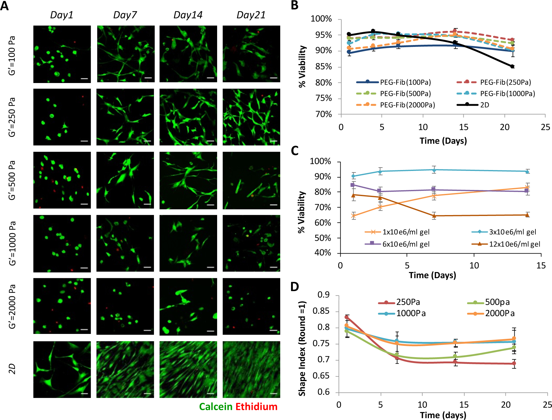
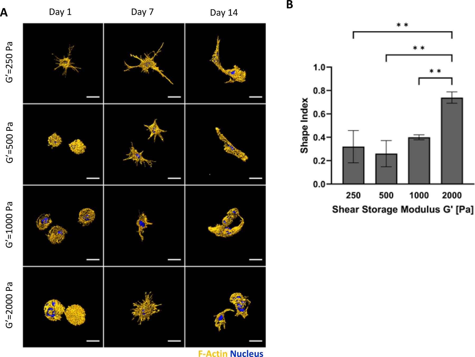
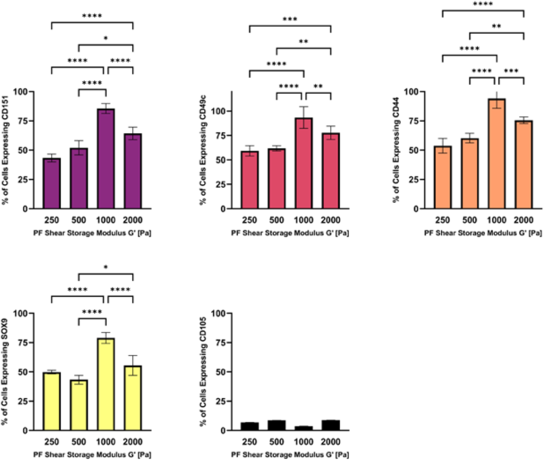
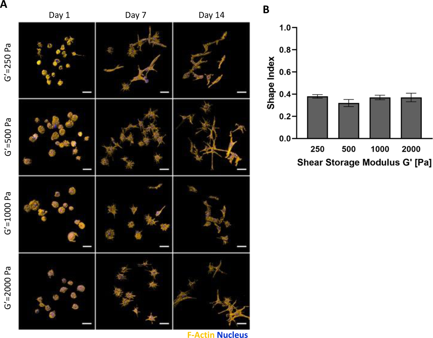
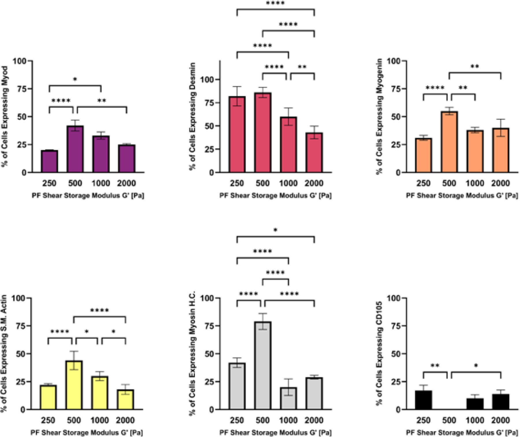
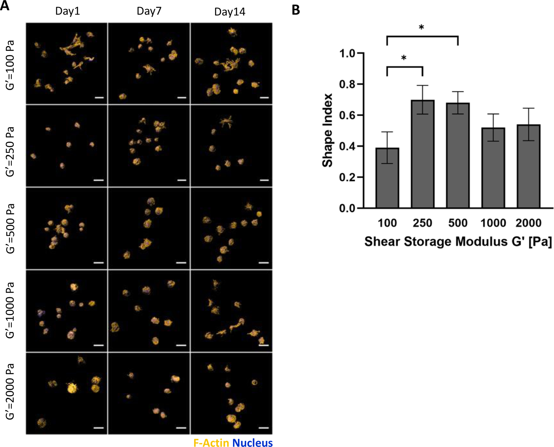
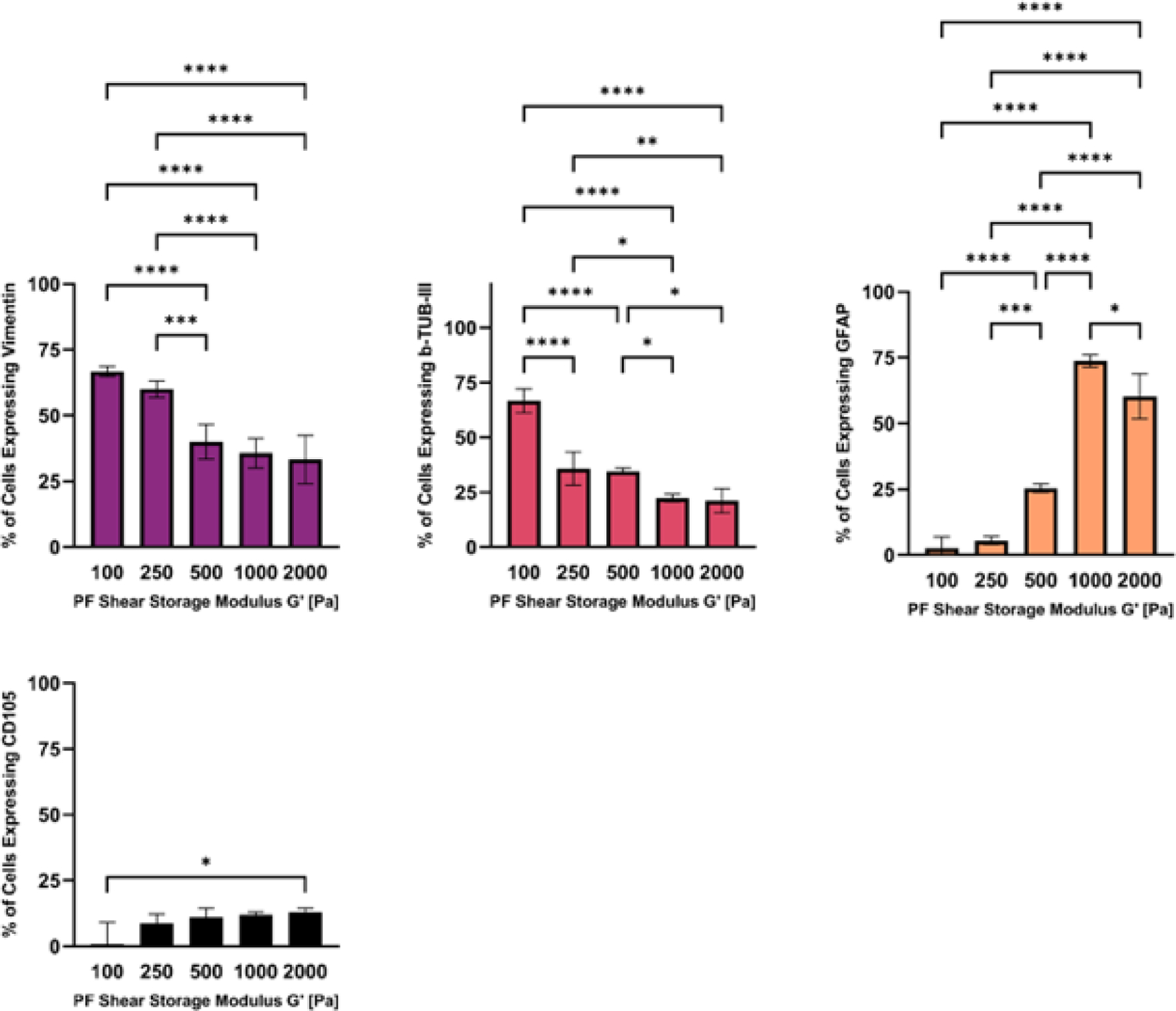
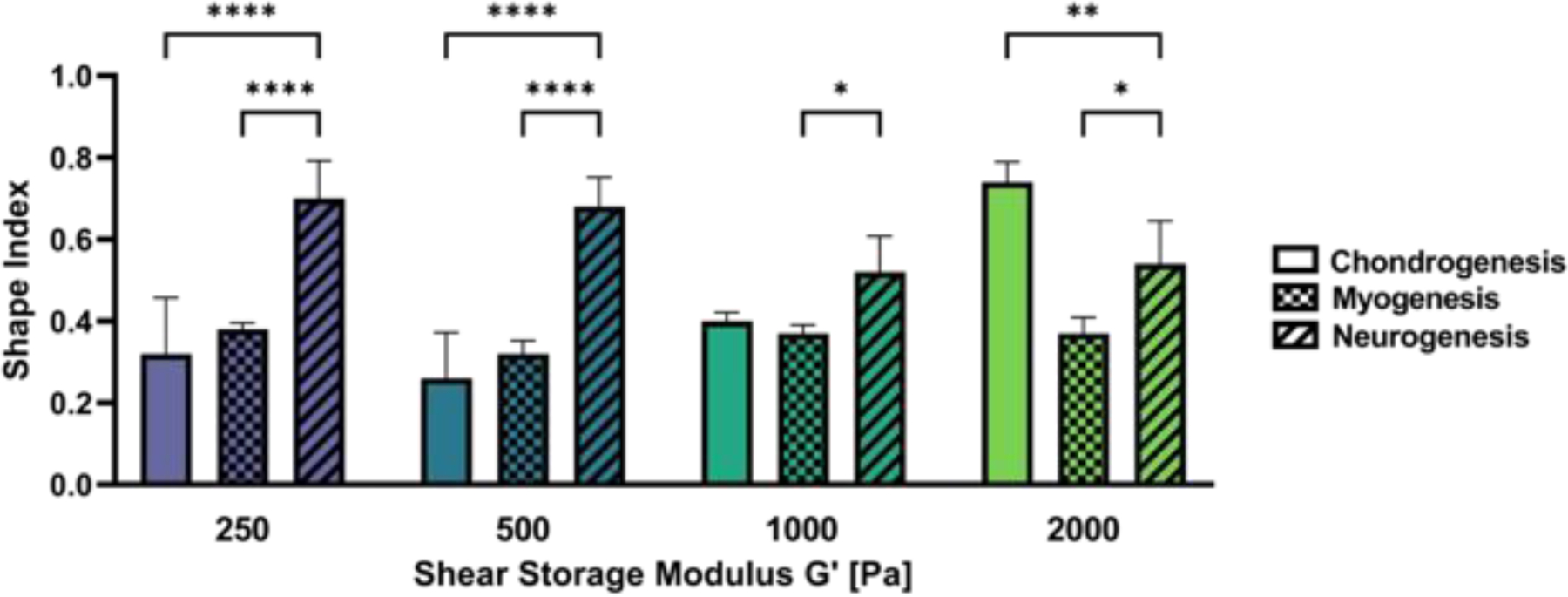
Human mesenchymal stromal cells (hMSCs) are of significant interest as a renewable source of therapeutically useful cells. In tissue engineering, hMSCs are implanted within a scaffold to provide enhanced capacity for tissue repair. The present study evaluates how mechanical properties of that scaffold can alter the phenotype and genotype of the cells, with the aim of augmenting hMSC differentiation along the myogenic, neurogenic or chondrogenic linages. The hMSCs were grown three-dimensionally (3D) in a hydrogel comprised of poly(ethylene glycol) (PEG)-conjugated to fibrinogen. The hydrogel's shear storage modulus (G'), which was controlled by increasing the amount of PEG-diacrylate cross-linker in the matrix, was varied in the range of 100-2000 Pascal (Pa). The differentiation into each lineage was initiated by a defined culture medium, and the hMSCs grown in the different modulus hydrogels were characterized using gene and protein expression. Materials having lower storage moduli (G' = 100 Pa) exhibited more hMSCs differentiating to neurogenic lineages. Myogenesis was favored in materials having intermediate modulus values (G' = 500 Pa), whereas chondrogenesis was favored in materials with a higher modulus (G' = 1000 Pa). Enhancing the differentiation pathway of hMSCs in 3D hydrogel scaffolds using simple modifications to mechanical properties represents an important achievement toward the effective application of these cells in tissue engineering.

Keywords: biomaterials; hydrogel; shear modulus; stem cells; tissue engineering.


Figures

Figure 1:
Figure 1:
Experimental setup used to identify modulus-dependent differentiation patterns of hMSCs in 3D PEG-fibrinogen (PF) hydrogel cultures. PF hydrogels are formed with hMSCs and 8 mg/mL PF hydrogel liquid precursor, cross-linked by photopolymerization, and cultivated for up to 14 days in bioreactors using culture medium for expansion or differentiation. Cell-laden hydrogels were recovered from the bioreactors at set time-points and analyzed for viability, morphometrics and differentiation.
Figure 2.
Figure 2.
Modulus-dependent viability of hMSCs in 3D-PF hydrogels. (A) Live-dead staining of hMSCs in PEG-fibrinogen hydrogels (3×10⁶ cells/ml) grown in expansion medium for up to 21 days; the modulus of the hydrogels did not affect the viability of the cells (scale bar=25μm). (B) Quantitative viability with DNA staining using propidium iodide (PI) confirms the qualitative results of hMSCs seeded in the different modulus PEG-Fib hydrogels. Cells grown on tissue culture plastic dishes (2D) were used as controls. (C) The effect of cell seeding density on hMSC viability was assessed in PF hydrogels with a G’=250Pa modulus, grown in expansion medium for up to 14 days. The highest percent of viable cells was evident in hydrogels made with a hMSC concentration of 3×10⁶cells/ml. The viability data is presented as the average plus/minus standard deviation; a significant difference between the treatments was observed by 2-way ANOVA (p < 0.05) (D) The morphology of hMSCs in the 3D-PF hydrogels was quantitatively evaluated for hMSCs in PEG-fibrinogen hydrogels (3×10⁶ cells/ml) grown in expansion medium for up to 21 days. The shape index (round=1) was calculated using the Imaris software; the initially rounded cells become more elongated over time as the cells spread three-dimensionally in the PF matrix. The shape index of the hMSCs was quantified as a function of hydrogel modulus to reveal a significant difference between the treatments (2-way ANOVA, p < 0.05).
Figure 3.
Figure 3.
Modulus-dependent morphometric analysis of hMSCs after 14 days in 3D PF hydrogels cultured in chondrogenic medium. (A) Representative confocal microscopy Imaris image segmentations of 3D hMSCs cultured in the 3D hydrogels. The cells are stained with an actin stain (orange) and a nucleus counterstain (Blue); Scale bar=25μm. (B) The shape index of the hMSCs was quantified as a function of modulus, indicating a correlation between cellular extensions and storage shear modulus (G′) after 14 days. The shape index (round=1) was calculated using the Imaris software. Data is presented as the average plus/minus standard deviation. **Indicates statistically significant differences between the modulus treatments (p < 0.01).
Figure 4.
Figure 4.
The modulus-dependent chondrogenic differentiation of hMSCs in 3D PF hydrogels was evaluated after 14 days in chondrogenic medium. Specific markers measured by flow cytometry include CD151, CD49c, CD44, Sox9 and CD105 (negative marker). Data is presented as the average plus/minus standard deviation. Statistically significant differences between the modulus treatments are indicated on the graphs: *p < 0.05; **p < 0.01; ***p < 0.001; ****p < 0.0001.
Figure 5.
Figure 5.
Modulus-dependent morphometric analysis of hMSCs after 14 days in 3D PF hydrogels cultured in myogenic medium. (A) Representative confocal microscopy Imaris image segmentations of 3D hMSCs cultured in the 3D hydrogels. The cells are stained with an actin stain (orange) and a nucleus counterstain (Blue); Scale bar=25μm. (B) The shape index of the hMSCs was quantified as a function of modulus, indicating a correlation between cellular extensions and storage shear modulus (G′) after 14 days. The shape index (round=1) was calculated using the Imaris software. Data is presented as the average plus/minus standard deviation. Statistically significant differences between the modulus treatments were not observed (p>0.05).
Figure 6:
Figure 6:
The modulus-dependent myogenic differentiation of hMSCs was evaluated after 14 days in 3D PF hydrogels cultured in myogenic medium. Specific markers measured by flow cytometry include MyoD, Desmin, Myogenin, S.M.Actin, Myosin H.C. and CD105 (negative marker). Data is presented as the average plus/minus standard deviation. Statistically significant differences between the modulus treatments are indicated on the graphs: *p < 0.05; **p < 0.01; ***p < 0.001; ****p < 0.0001.
Figure 7.
Figure 7.
Modulus-dependent morphometric analysis of hMSCs after 14 days in 3D PF hydrogels cultured in neurogenic medium. (A) Representative confocal microscopy Imaris image segmentations of 3D hMSCs cultured in the 3D hydrogels. The cells are stained with an actin stain (orange) and a nucleus counterstain (Blue); Scale bar=25μm. (B) The shape index of the hMSCs was quantified as a function of modulus, indicating a correlation between cellular extensions and storage shear modulus (G′) after 14 days. The shape index (round=1) was calculated using the Imaris software. Data is presented as the average plus/minus standard deviation. *Indicates statistically significant differences between the modulus treatments (p < 0.05).
Figure 8.
Figure 8.
The modulus-dependent neurogenic differentiation of hMSCs after 14 days in 3D PF hydrogels cultured in neurogenic medium. Specific markers measured by flow cytometry include the pre-neural marker Vimentin, neural marker β-Tubulin-III, glial marker GFAP, and CD105 (negative marker). Data is presented as the average plus/minus standard deviation. Statistically significant differences between the modulus treatments are indicated on the graphs: *p < 0.05; **p < 0.01; ***p < 0.001; ****p < 0.0001.
Figure 9:
Figure 9:
Modulus-dependent morphometric analysis of hMSCs after 14 days in 3D PF hydrogels cultured in chondrogenic, myogenic and neurogenic medium. The differentiation conditions at any given modulus treatment have a significant impact on the cell morphology. Statistically significant differences between the treatments are indicated on the graph: *p < 0.05; **p < 0.01; ***p < 0.001; ****p < 0.0001.

Similar articles

Cited by

References

    1. Caddeo S, Boffito M, Sartori S. Tissue engineering Approaches in the Design of Healthy and Pathological In Vitro Tissue Models. Frontiers in Bioengineering and Biotechnology 2017;5. - PMC - PubMed
    1. Lev R, Seliktar D. Hydrogel biomaterials and their therapeutic potential for muscle injuries and muscular dystrophies. Journal of the Royal Society Interface 2018;15(138). - PMC - PubMed
    1. Berdichevski A, Yameen HS, Dafni H, Neeman M, Seliktar D. Using bimodal MRI/fluorescence imaging to identify host angiogenic response to implants. Proceedings of the National Academy of Sciences of the United States of America 2015;112(16):5147–5152. - PMC - PubMed
    1. Ciocci M, Cacciotti I, Seliktar D, Melino S. Injectable silk fibroin hydrogels functionalized with microspheres as adult stem cells-carrier systems. International Journal of Biological Macromolecules 2018;108:960–971. - PubMed
    1. Birman T, Seliktar D. Injectability of Biosynthetic Hydrogels: Consideration for Minimally Invasive Surgical Procedures and 3D Bioprinting. Advanced Functional Materials 2021;31(29).

Publication types