

Diagnostic Challenges of Neuromuscular Disorders after Whole Exome Sequencing
- PMID: 37066920
- PMCID: PMC10357215
- DOI: 10.3233/JND-230013
Diagnostic Challenges of Neuromuscular Disorders after Whole Exome Sequencing
Abstract
Background: Whole-exome sequencing (WES) facilitates the diagnosis of hereditary neuromuscular disorders. To achieve an accurate diagnosis, physicians should interpret the genetic report carefully along with clinical information and examinations. We described our experience with (1) clinical validation in patients with variants found using WES and (2) a diagnostic approach for those with negative findings from WES.
Methods: WES was performed on patients with the clinical impression of hereditary neuromuscular disorders. Information on clinical manifestations, neurological examination, electrodiagnostic studies, histopathology of muscle and nerve, and laboratory tests were collected.
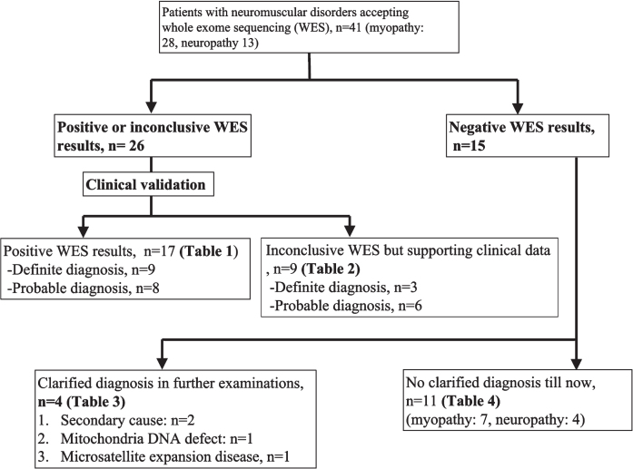
Results: Forty-one patients (Male/Female: 18/23, age of onset: 34.5±15.9) accepted WES and were categorized into four scenarios: (1) patients with a positive WES result, (2) patients with an inconclusive WES result but supporting clinical data, (3) negative findings from WES, but a final diagnosis after further work-up, and (4) undetermined etiology from WES and in further work-ups. The yield rate of the initial WES was 63.4% (26/41). Among these, seventeen patients had positive WES result, while the other nine patients had inconclusive WES result but supporting clinical data. Notably, in the fifteen patients with negative findings from WES, four patients (26.7%) achieved a diagnosis after further workup: tumor-induced osteomalacia, metabolic myopathy with pathogenic variants in mitochondrial DNA, microsatellite expansion disease, and vasculitis-related neuropathy. The etiologies remained undetermined in eleven patients (myopathy: 7, neuropathy: 4) after WES and further workup.
Conclusions: It is essential to design genotype-guided molecular studies to correlate the identified variants with their clinical features. For patients who had negative findings from WES, acquired diseases, mitochondrial DNA disorders and microsatellite expansion diseases should be considered.
Keywords: Whole-exome sequencing; motor neuron disease; myopathy; neuromuscular disorders; neuropathy.
Conflict of interest statement
None.
Figures
Similar articles
-
Diagnostic value of whole-exome sequencing in Chinese pediatric-onset neuromuscular patients.Mol Genet Genomic Med. 2020 May;8(5):e1205. doi: 10.1002/mgg3.1205. Epub 2020 Mar 10. Mol Genet Genomic Med. 2020. PMID: 32154989 Free PMC article.
-
Panel-Based Exome Sequencing for Neuromuscular Disorders as a Diagnostic Service.J Neuromuscul Dis. 2019;6(2):241-258. doi: 10.3233/JND-180376. J Neuromuscul Dis. 2019. PMID: 31127727
-
Molecular findings in patients for whole exome sequencing and mitochondrial genome assessment.Clin Chim Acta. 2024 Jul 15;561:119774. doi: 10.1016/j.cca.2024.119774. Epub 2024 Jun 8. Clin Chim Acta. 2024. PMID: 38852791
-
Utility of exome sequencing for the diagnosis of pediatric-onset neuromuscular diseases beyond diagnostic yield: a narrative review.Neurol Sci. 2024 Apr;45(4):1455-1464. doi: 10.1007/s10072-023-07210-z. Epub 2023 Nov 22. Neurol Sci. 2024. PMID: 37989827 Free PMC article. Review.
-
[Exome diagnostics in neurology].Nervenarzt. 2019 Feb;90(2):131-137. doi: 10.1007/s00115-018-0667-1. Nervenarzt. 2019. PMID: 30645660 Review. German.
Cited by
-
Concordance Between Biochemical and Molecular Diagnosis Obtained by WES in Mexican Patients with Inborn Errors of Intermediary Metabolism: Utility for Therapeutic Management.Int J Mol Sci. 2024 Oct 31;25(21):11722. doi: 10.3390/ijms252111722. Int J Mol Sci. 2024. PMID: 39519275 Free PMC article.
-
Comparative genetic diagnostic evaluation of pediatric neuromuscular diseases in a consanguineous population.Sci Rep. 2025 Jan 2;15(1):231. doi: 10.1038/s41598-024-81744-w. Sci Rep. 2025. PMID: 39747233 Free PMC article.
-
Two distinct phenotypes and a novel mutation in limb-girdle muscular dystrophy R7 telethonin-related patients from Thai neuromuscular center.Neurol Sci. 2025 Aug;46(8):3929-3940. doi: 10.1007/s10072-025-08158-y. Epub 2025 Apr 7. Neurol Sci. 2025. PMID: 40195250
-
NEUROMYODredger: Whole Exome Sequencing for the Diagnosis of Neurodevelopmental and Neuromuscular Disorders in Seven Countries.Clin Genet. 2025 Feb 25;108(3):318-22. doi: 10.1111/cge.14736. Online ahead of print. Clin Genet. 2025. PMID: 40000157 Free PMC article.
-
Inherited Metabolic Diseases from Past to Present: A Bibliometric Analysis (1968-2023).Children (Basel). 2023 Jul 12;10(7):1205. doi: 10.3390/children10071205. Children (Basel). 2023. PMID: 37508702 Free PMC article.
References
-
- Westra D, et al.. Panel-based exome sequencing for neuromuscular disorders as a diagnostic service. J Neuromuscul Dis. 2019;6(2):241–58. - PubMed
-
- Ababneh NA, et al.. The utility of whole-exome sequencing in accurate diagnosis of neuromuscular disorders in consanguineous families in Jordan. Clin Chim Acta. 2021;523:330–8. - PubMed
-
- Hartley T, et al.. Whole-exome sequencing is a valuable diagnostic tool for inherited peripheral neuropathies: Outcomes from a cohort of 50 families. Clin Genet. 2018;93(2):301–9. - PubMed
MeSH terms
Substances
LinkOut - more resources
Full Text Sources
Medical
