. 2023 Jul 5;21(7):698-712.
doi: 10.1158/1541-7786.MCR-23-0003.

BZW2 Inhibition Reduces Colorectal Cancer Growth and Metastasis

Affiliations

BZW2 Inhibition Reduces Colorectal Cancer Growth and Metastasis

Sumit Agarwal et al. Mol Cancer Res. .

Abstract

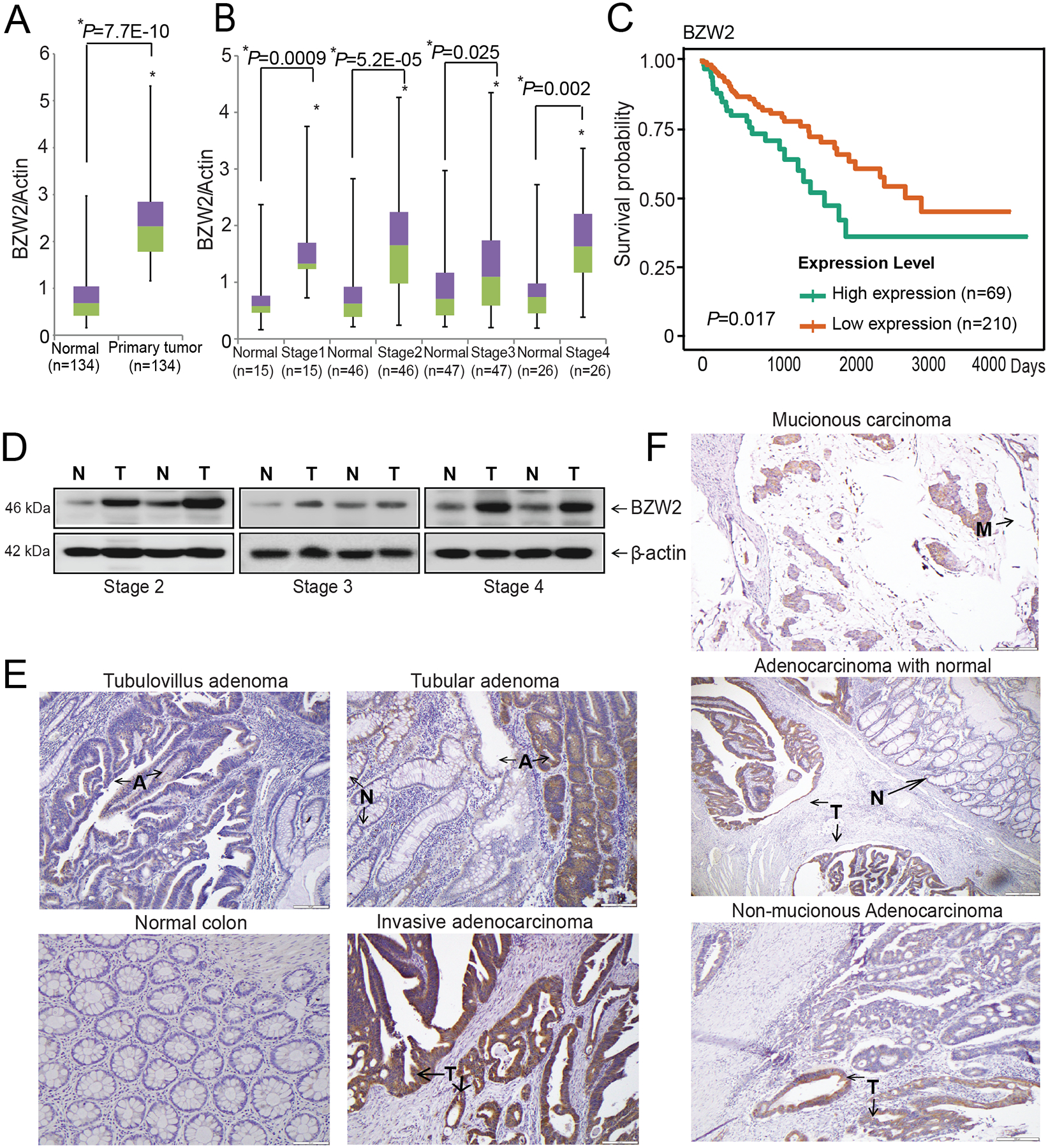
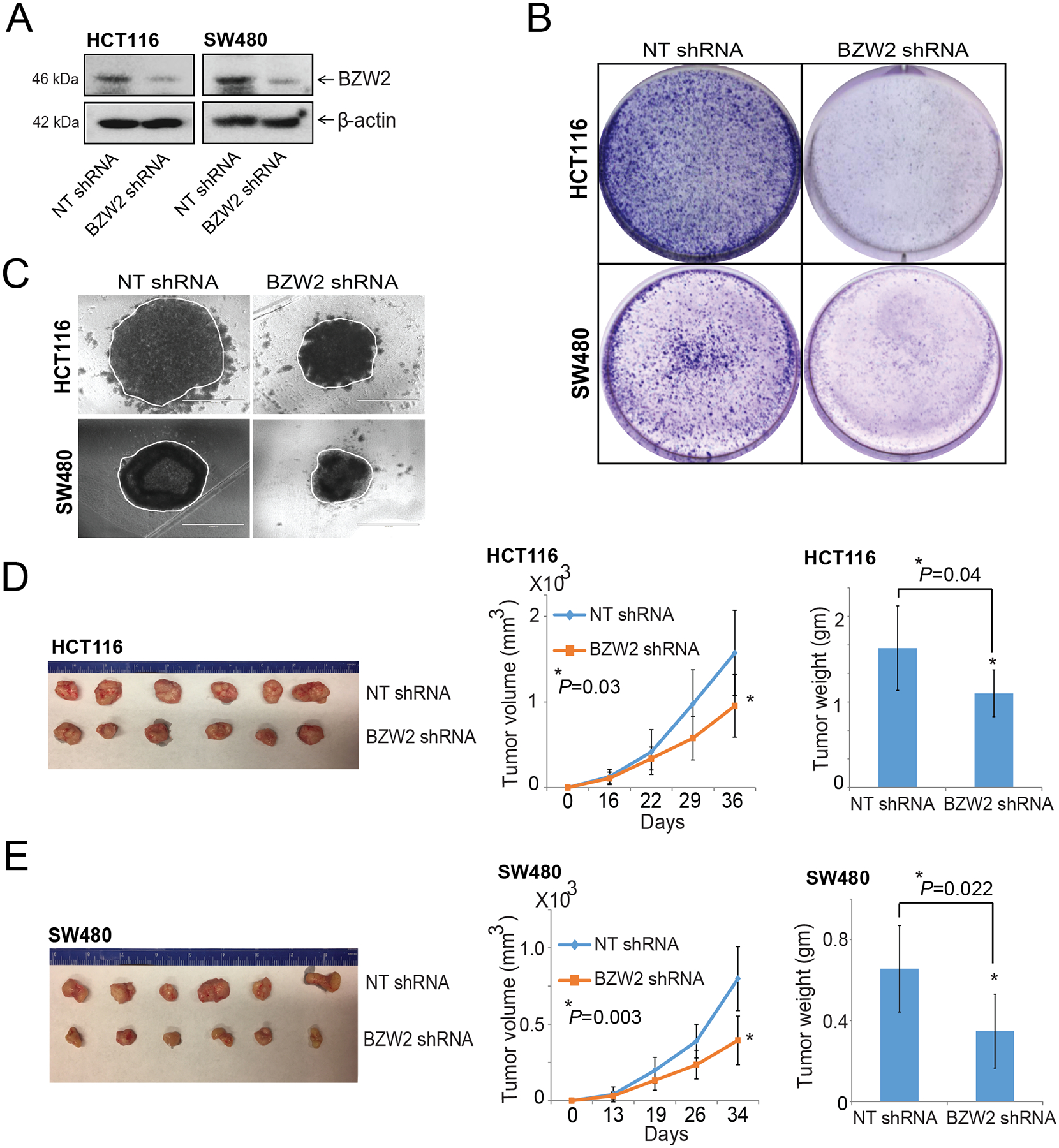
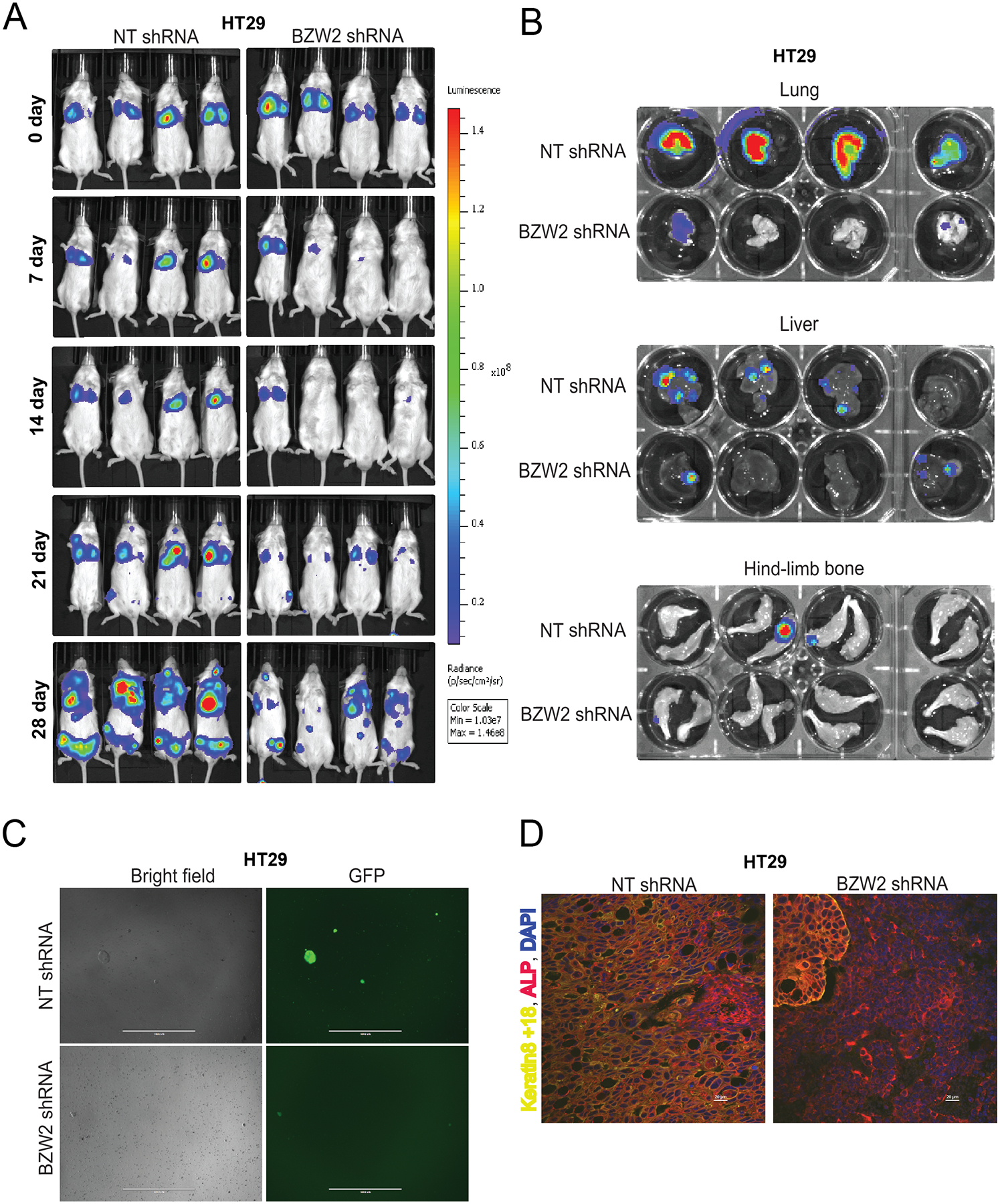
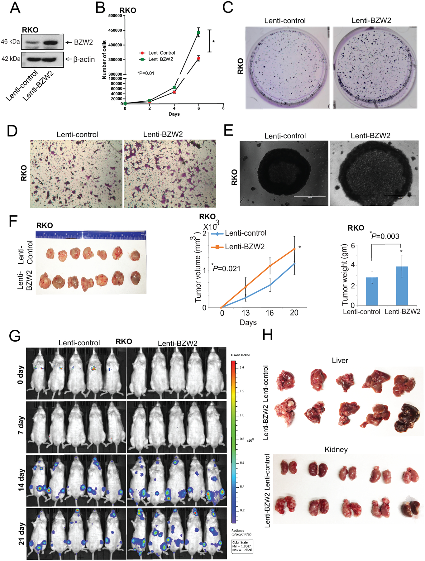
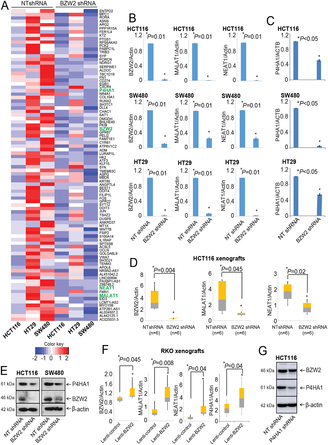
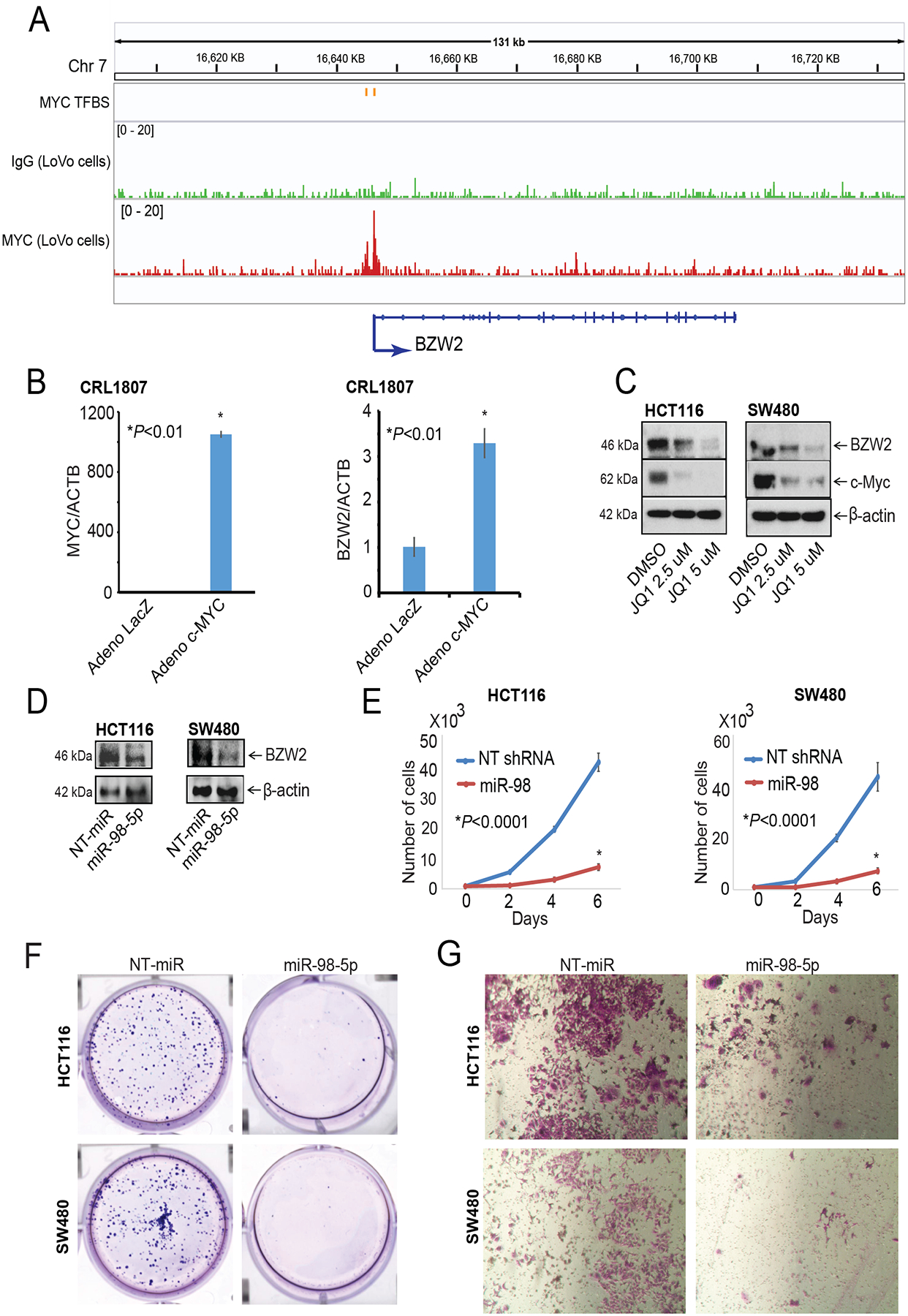
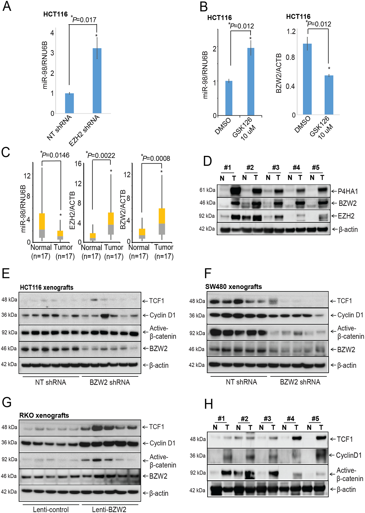
Because survival of patients with metastatic colorectal cancer remain poor, there is an urgent need to identify potential novel druggable targets that are associated with colorectal cancer progression. One such target, basic leucine zipper and W2 domains 2 (BZW2), is involved in regulation of protein translation, and its overexpression is associated with human malignancy. Thus, we investigated the expression and regulation of BZW2, assessed its role in activation of WNT/β-catenin signaling, identified its downstream molecules, and demonstrated its involvement in metastasis of colorectal cancer. In human colorectal cancers, high mRNA and protein expression levels of BZW2 were associated with tumor progression. BZW2-knockdown reduced malignant phenotypes, including cell proliferation, invasion, and spheroid and colony formation. BZW2-knockdown also reduced tumor growth and metastasis; conversely, transfection of BZW2 into BZW2 low-expressing colorectal cancer cells promoted malignant features, including tumor growth and metastasis. BZW2 expression was coordinately regulated by microRNA-98, c-Myc, and histone methyltransferase enhancer of zeste homolog 2 (EZH2). RNA sequencing analyses of colorectal cancer cells modulated for BZW2 identified P4HA1 and the long noncoding RNAs, MALAT1 and NEAT1, as its downstream targets. Further, BZW2 activated the Wnt/β-catenin signaling pathway in colorectal cancers expressing wild-type β-catenin. In sum, our study suggests the possibility of targeting BZW2 expression by inhibiting EZH2 and/or c-Myc.

Implications: FDA-approved small-molecule inhibitors of EZH2 can indirectly target BZW2 and because BZW2 functions as an oncogene, these inhibitors could serve as therapeutic agents for colorectal cancer.


Conflict of interest statement

Conflict of interest: The authors declare no conflict of interest.

Figures

Figure 1.
Figure 1.
BZW2 upregulation in CRC. A, BZW2 RNA expression in frozen CRC tissues of whole cohort with matched normals was quantified by qRT-PCR using specific primers. Histograph presenting group comparisons of relative BZW2 RNA expression in frozen paired normal (n=134) and tumor tissues (n=134) of CRC patients. B, In-house validation qRT-PCR analysis showing BZW2 expression in various pathologic stages (Stage 1, n=15; Stage 2, n=46; Stage 3, n=47; and Stage 4, n=26). C, Kaplan-Meier curves for CRC-specific survival with low and high BZW2 expression according to data from TCGA (log rank, P=0.017). D, Western blot analyses showing BZW2 levels in frozen CRCs and in paired adjacent non-cancerous tissues with respect to the stage of cancer. E & F, IHC staining showing the BZW2 expression in adjacent non-tumor tissue (denoted by N), tubule-villus adenoma, tubular adenoma (denoted by A), mucinous carcinoma (denoted by M), adenocarcinoma, and invasive CRC tissues. Hematoxylin was used for nuclear staining. Scale bar, 100 μm.
Figure 2.
Figure 2.
BZW2 involvement in the proliferation, spheroid formation, and tumor growth of CRC cells. A, The expression of BZW2 was measured by Western blots after BZW2-knockdown in CRC cells, HCT116 and SW480. β-Actin was used as a loading control. B, Representative photographs of the colony formation assay for NT shRNA and BZW2-knockdown CRC cells. C, Phase-contrast microscopy images of CRC spheroids of NT shRNA and BZW2-knockdown cells (scale bar, 1000 μm). NSG mice were injected subcutaneously with HCT116 or SW480 cells (1.0×10⁶/mouse) manifesting various TP53 status with NT shRNA or BZW2 shRNA. D, Representative images of tumors at the end of the experiment showing HCT116, and E, SW480 xenograft tumors exhibiting control NT shRNA and BZW2 shRNA. Gross observation of BZW2 shRNA xenografts showing smaller tumors for CRC cells as compared to those of NT shRNA xenografts. Tumor volumes and tumor weights were measured and plotted as means ± SD.
Figure 3.
Figure 3.
BZW2 involvement in CRC metastasis in a tail-vein metastasis model. A, HT29 cells exhibiting BZW2 shRNA or NT shRNA were injected into tail veins of NSG mice, and bioluminescence imaging was performed weekly by injecting luciferin intraperitoneally. B, Ex vivo luminescence of organs procured after sacrifice of mice. C, Cultures of mouse bone marrow in media containing puromycin to select cancer cells. Representative phase-contrast and GFP images after 7 days showing cultured CRC cells from bone marrows of mice injected with HT29 cells exhibiting stably expressed NT shRNA or BZW2 shRNA (scale bar, 1000 μm. D, Keratin 8+18 (yellow) and ALP (red) co-staining of bone lesions of NT shRNA or BZW2 shRNA cells. DAPI was used for nuclear staining (scale bar, 20 μm).
Figure 4.
Figure 4.
Ectopic expression of BZW2 enhances malignant phenotypes of CRC. A, Expression of BZW2 in RKO cells was analyzed by Western blotting. B, Cell proliferation assay evaluating the effect of BZW2 overexpression on proliferation of RKO cells. C, Representative images of crystal violet-stained colonies of RKO cells transfected with a lenti-control or lenti-BZW2. D, Invasion capacity of BZW2-overexpressing or control vector RKO cells examined by a Transwell Matrigel invasion assay. E, Representative spheroids formed after BZW2 overexpression in RKO cells. F, BZW2 overexpressing or control vector RKO cells (2 ×10⁶/mouse) were injected subcutaneously into NSG mice. On day 20, the six mice were sacrificed, and tumors were harvested. Values are presented as the means ± SD. G, In vivo luminescence imaging of NSG mice with RKO cells exhibiting BZW2 overexpression or control vector was performed. H, Representative photographs of liver and kidney retrieved from the metastasis experiment.
Figure 5.
Figure 5.
MALAT1 and NEAT1 as downstream targets of BZW2 in CRC. A, Heatmap of differentially expressed genes from expression profiling of stable BZW2-knockdown HCT116, SW480, and HT29 cells. B, qRT PCR analysis of BZW2, MALAT1, and NEAT1 and C, P4HA1 in RNA of HCT116, SW480, and HT29 cells with stable BZW2-knockdown. D, RNA expression of BZW2, MALAT1, and NEAT1 in xenografts of HCT116 exhibiting BZW2 knockdown. E, Western blot analysis showing the expression of P4HA1 in BZW2-knockdown CRC cells. F, RNA expression of BZW2, MALAT1, NEAT1, and P4HA1 in BZW2-overexpressing RKO xenografts. Quantitative data are means ± SD. *P < 0.01. G, The expression of BZW2 was measured after knockdown of P4HA1.
Figure 6.
Figure 6.
BZW2 regulation by MYC and miR-98 in CRC. A, MYC transcription binding sites in the BZW2 promoter region: IGV snapshots of MYC ChIP-seq data [GSE51290] for gene and promoter region of BZW2. Top track shows MYC binding sites; the next two tracks indicate ChIP-seq read coverage on antibody treatment (MYC) and IgG in CRC cells (LoVo). LacZ control and MYC adenovirus were transduced into SV-40 transformed CRL1807 colon cells. B qRT-PCR analysis was performed to assess the RNA expression of MYC and BZW2 after MYC overexpression. C, HCT116, and SW480 cells were exposed to JQ1 followed by probing for BZW2 and c-Myc. D, HCT116, and SW480 cells were transfected with NT-pre-miR or miR-98. Immunoblot analyses were performed to assess BZW2 protein expression. Ectopic expression of NT-pre-miR or miR-98 was performed for HCT116, and SW480 cells to assess E, cell proliferation, F, colony formation, and G, invasion.
Figure 7.
Figure 7.
EZH2 regulates BZW2, which activates the WNT/β-catenin signaling pathway in CRC. A, qRT-PCR analysis showing the expression of miR-98 and B, qRT-PCR analysis to assess the expressions of miR-98 and BZW2 in GSK-126-treated CRC cells. C, qRT-PCR analysis showing the relation of miR-98 with EZW2 or BZW2 in human CRC samples. D, Protein levels of EZH2, BZW2, and P4HA1 were measured in human CRCs (denoted by T) and matched adjacent normal breast (denoted by N) tissues (n = 5) by Western blot analysis. WNT/β-catenin signaling molecules, active-β-catenin, LEF1, and TCF1 protein levels were measured in xenografts of E, HCT116. F, SW480 and G, RKO having modulated BZW2 expression. H, Protein levels of WNT/β-catenin signaling molecules were measured in CRC (denoted by T) and matched adjacent normal (denoted by N) tissues (n = 5), that were used in Fig 7D, by Western blot analysis. Quantitative data are presented in means ± SD. *P < 0.01.

Similar articles

Cited by

References

    1. Siegel RL, Miller KD, Goding Sauer A, Fedewa SA, Butterly LF, Anderson JC, et al. Colorectal cancer statistics, 2021. CA Cancer J Clin. 2021. - PubMed
    1. Singh CR, Watanabe R, Zhou D, Jennings MD, Fukao A, Lee B, et al. Mechanisms of translational regulation by a human eIF5-mimic protein. Nucleic Acids Res. 2011;39(19):8314–28. - PMC - PubMed
    1. Cheng DD, Li SJ, Zhu B, Yuan T, Yang QC, Fan CY. Downregulation of BZW2 inhibits osteosarcoma cell growth by inactivating the Akt/mTOR signaling pathway. Oncol Rep. 2017;38(4):2116–22. - PMC - PubMed
    1. Jin X, Liao M, Zhang L, Yang M, Zhao J. Role of the novel gene BZW2 in the development of hepatocellular carcinoma. J Cell Physiol. 2019. - PubMed
    1. Gao H, Yu G, Zhang X, Yu S, Sun Y, Li Y. BZW2 gene knockdown induces cell growth inhibition, G1 arrest and apoptosis in muscle-invasive bladder cancers: A microarray pathway analysis. J Cell Mol Med. 2019;23(6):3905–15. - PMC - PubMed

Publication types

MeSH terms