

Genome-Wide Mapping of the Escherichia coli PhoB Regulon Reveals Many Transcriptionally Inert, Intragenic Binding Sites
- PMID: 37067422
- PMCID: PMC10294691
- DOI: 10.1128/mbio.02535-22
Genome-Wide Mapping of the Escherichia coli PhoB Regulon Reveals Many Transcriptionally Inert, Intragenic Binding Sites
Abstract
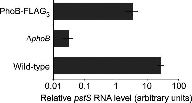
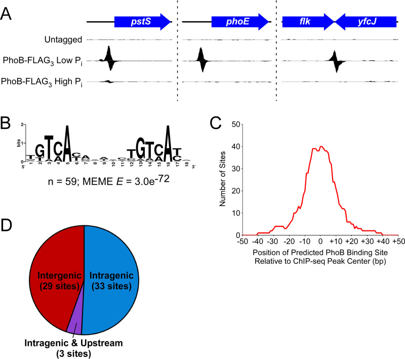
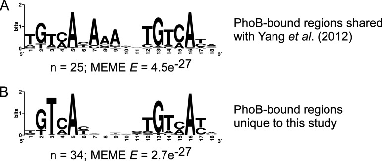
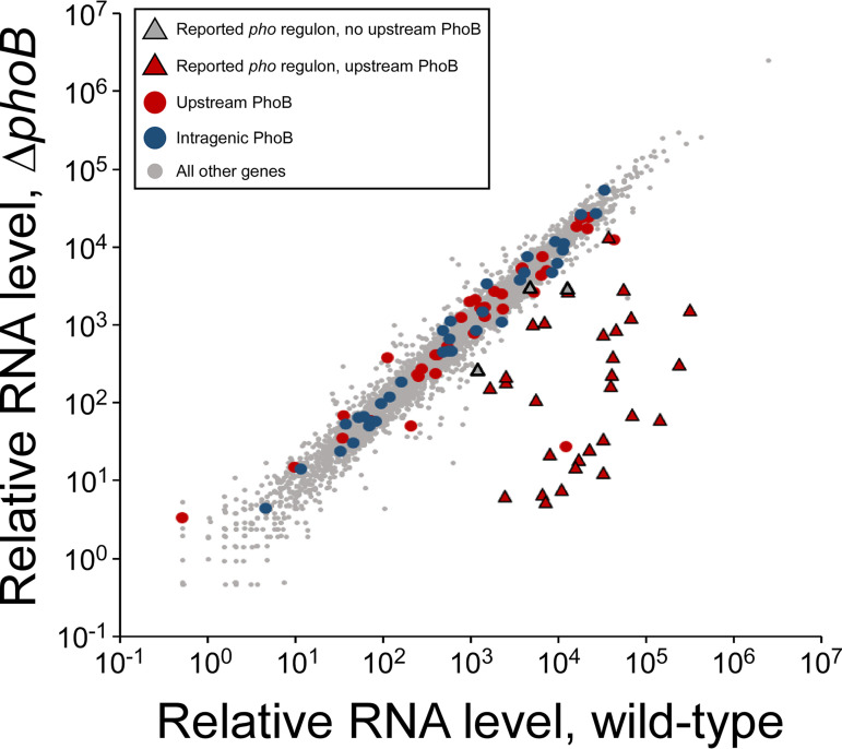
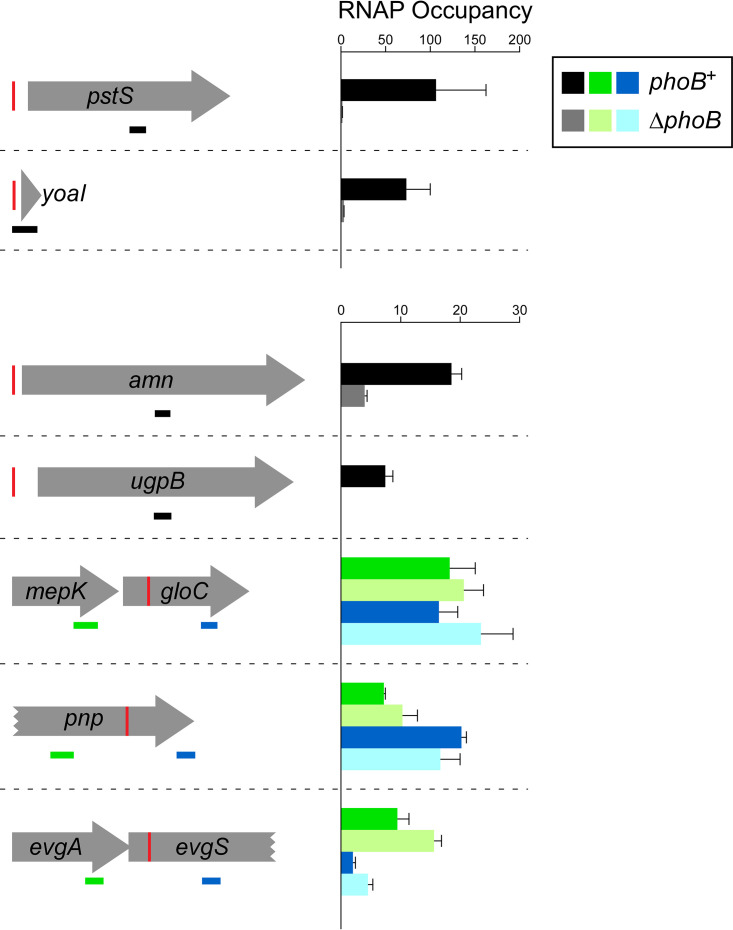
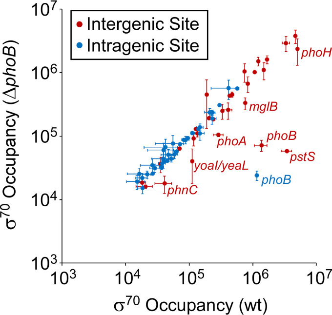
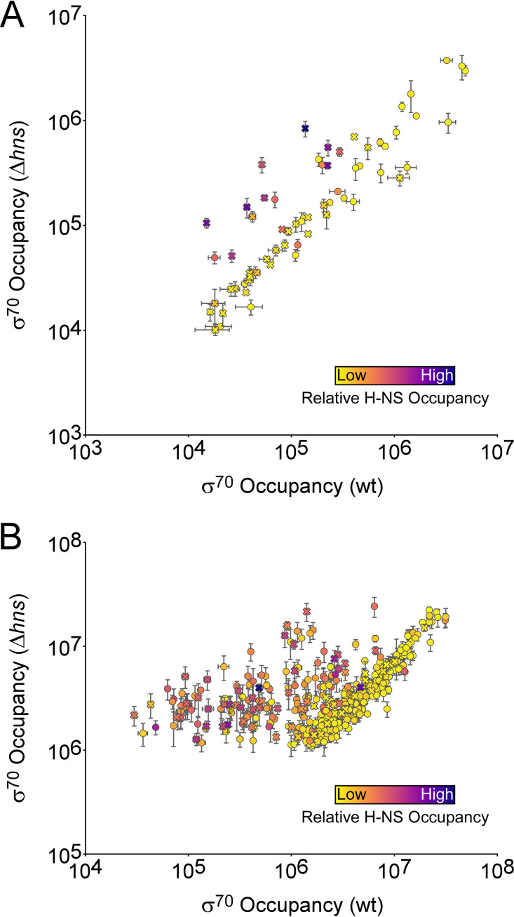
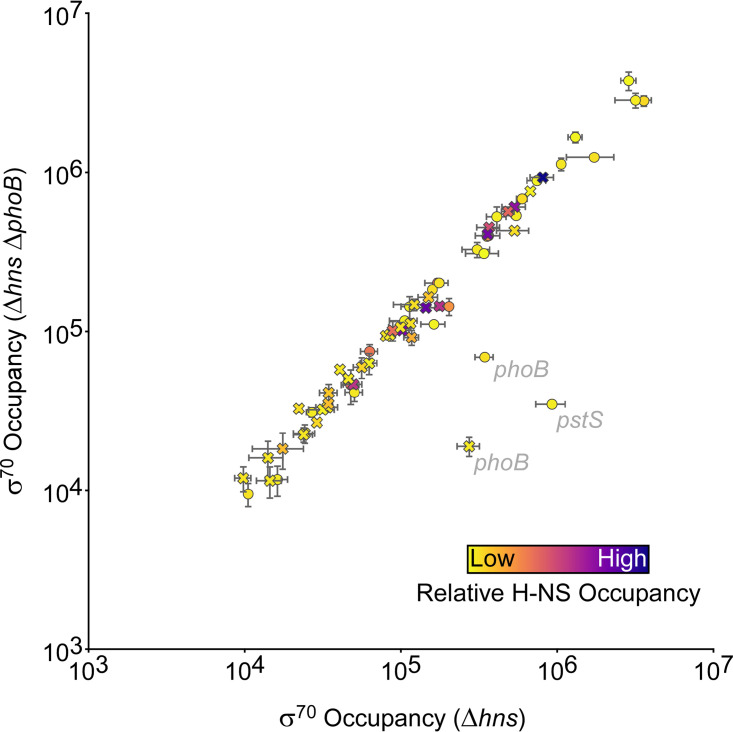
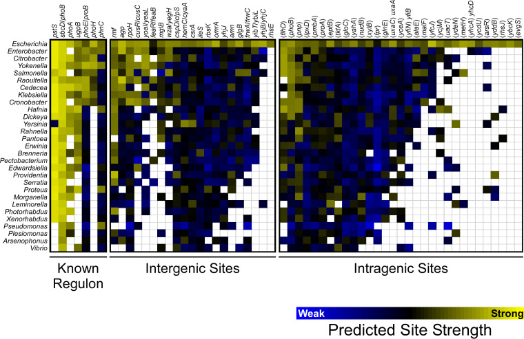
Genome-scale analyses have revealed many transcription factor binding sites within, rather than upstream of, genes, raising questions as to the function of these binding sites. Here, we use complementary approaches to map the regulon of the Escherichia coli transcription factor PhoB, a response regulator that controls transcription of genes involved in phosphate homeostasis. Strikingly, the majority of PhoB binding sites are located within genes, but these intragenic sites are not associated with detectable transcription regulation and are not evolutionarily conserved. Many intragenic PhoB sites are located in regions bound by H-NS, likely due to shared sequence preferences of PhoB and H-NS. However, these PhoB binding sites are not associated with transcription regulation even in the absence of H-NS. We propose that for many transcription factors, including PhoB, binding sites not associated with promoter sequences are transcriptionally inert and hence are tolerated as genomic "noise." IMPORTANCE Recent studies have revealed large numbers of transcription factor binding sites within the genes of bacteria. The function, if any, of the vast majority of these binding sites has not been investigated. Here, we map the binding of the transcription factor PhoB across the Escherichia coli genome, revealing that the majority of PhoB binding sites are within genes. We show that PhoB binding sites within genes are not associated with regulation of the overlapping genes. Indeed, our data suggest that bacteria tolerate the presence of large numbers of nonregulatory, intragenic binding sites for transcription factors and that these binding sites are not under selective pressure.
Keywords: ChIP-seq; PhoB; pho regulon; transcription factors.
Conflict of interest statement
The authors declare no conflict of interest.
Figures
Update of
-
Genome-wide mapping of the Escherichia coli PhoB regulon reveals many transcriptionally inert, intragenic binding sites.bioRxiv [Preprint]. 2023 Feb 7:2023.02.07.527549. doi: 10.1101/2023.02.07.527549. bioRxiv. 2023. Update in: mBio. 2023 Jun 27;14(3):e0253522. doi: 10.1128/mbio.02535-22. PMID: 36798257 Free PMC article. Updated. Preprint.
Similar articles
-
Genome-wide mapping of the Escherichia coli PhoB regulon reveals many transcriptionally inert, intragenic binding sites.bioRxiv [Preprint]. 2023 Feb 7:2023.02.07.527549. doi: 10.1101/2023.02.07.527549. bioRxiv. 2023. Update in: mBio. 2023 Jun 27;14(3):e0253522. doi: 10.1128/mbio.02535-22. PMID: 36798257 Free PMC article. Updated. Preprint.
-
Identification and characterization of novel phosphate regulon genes, ecs0540-ecs0544, in Escherichia coli O157:H7.Mol Genet Genomics. 2010 Sep;284(3):197-205. doi: 10.1007/s00438-010-0559-y. Epub 2010 Jul 17. Mol Genet Genomics. 2010. PMID: 20640580
-
Identification of the PhoB Regulon and Role of PhoU in the Phosphate Starvation Response of Caulobacter crescentus.J Bacteriol. 2015 Oct 19;198(1):187-200. doi: 10.1128/JB.00658-15. Print 2016 Jan 1. J Bacteriol. 2015. PMID: 26483520 Free PMC article.
-
Gene regulation by phosphate in enteric bacteria.J Cell Biochem. 1993 Jan;51(1):47-54. doi: 10.1002/jcb.240510110. J Cell Biochem. 1993. PMID: 8432742 Review.
-
The Pho regulon and the pathogenesis of Escherichia coli.Vet Microbiol. 2011 Nov 21;153(1-2):82-8. doi: 10.1016/j.vetmic.2011.05.043. Epub 2011 Jun 1. Vet Microbiol. 2011. PMID: 21700403 Review.
Cited by
-
Translation profiling of stress-induced small proteins reveals a novel link among signaling systems.bioRxiv [Preprint]. 2024 Oct 31:2024.09.13.612970. doi: 10.1101/2024.09.13.612970. bioRxiv. 2024. PMID: 39345582 Free PMC article. Preprint.
-
Phosphorus starvation response and PhoB-independent utilization of organic phosphate sources by Salmonella enterica.Microbiol Spectr. 2023 Dec 12;11(6):e0226023. doi: 10.1128/spectrum.02260-23. Epub 2023 Oct 3. Microbiol Spectr. 2023. PMID: 37787565 Free PMC article.
-
Phosphorus deficiency alleviates iron limitation in Synechocystis cyanobacteria through direct PhoB-mediated gene regulation.Nat Commun. 2024 May 24;15(1):4426. doi: 10.1038/s41467-024-48847-4. Nat Commun. 2024. PMID: 38789507 Free PMC article.
-
An intracellular phosphorus-starvation signal activates the PhoB/PhoR two-component system in Salmonella enterica.mBio. 2024 Sep 11;15(9):e0164224. doi: 10.1128/mbio.01642-24. Epub 2024 Aug 6. mBio. 2024. PMID: 39152718 Free PMC article.
-
Combined transcriptomic and ChIPseq analyses of the Bordetella pertussis RisA regulon.mSystems. 2024 Apr 16;9(4):e0095123. doi: 10.1128/msystems.00951-23. Epub 2024 Mar 12. mSystems. 2024. PMID: 38470037 Free PMC article.
References
-
- Galagan JE, Minch K, Peterson M, Lyubetskaya A, Azizi E, Sweet L, Gomes A, Rustad T, Dolganov G, Glotova I, Abeel T, Mahwinney C, Kennedy AD, Allard R, Brabant W, Krueger A, Jaini S, Honda B, Yu W-H, Hickey MJ, Zucker J, Garay C, Weiner B, Sisk P, Stolte C, Winkler JK, Van de Peer Y, Iazzetti P, Camacho D, Dreyfuss J, Liu Y, Dorhoi A, Mollenkopf H-J, Drogaris P, Lamontagne J, Zhou Y, Piquenot J, Park ST, Raman S, Kaufmann SHE, Mohney RP, Chelsky D, Moody DB, Sherman DR, Schoolnik GK. 2013. The Mycobacterium tuberculosis regulatory network and hypoxia. Nature 499:178–183. doi:10.1038/nature12337. - DOI - PMC - PubMed
-
- Knapp GS, Lyubetskaya A, Peterson MW, Gomes ALC, Ma Z, Galagan JE, McDonough KA. 2015. Role of intragenic binding of cAMP responsive protein (CRP) in regulation of the succinate dehydrogenase genes Rv0249c-Rv0247c in TB complex mycobacteria. Nucleic Acids Res 43:5377–5393. doi:10.1093/nar/gkv420. - DOI - PMC - PubMed
Publication types
MeSH terms
Substances
Grants and funding
LinkOut - more resources
Full Text Sources
Molecular Biology Databases
Miscellaneous
